6WUV

Crystal Structure of Recombinant Human Acetylcholinesterase Inhibited by GA


Experimental Data Snapshot

  • Method: X-RAY DIFFRACTION
  • Resolution: 2.63 Å
  • R-Value Free: 0.206 
  • R-Value Work: 0.170 
  • R-Value Observed: 0.172 

Starting Model: experimental
View more details

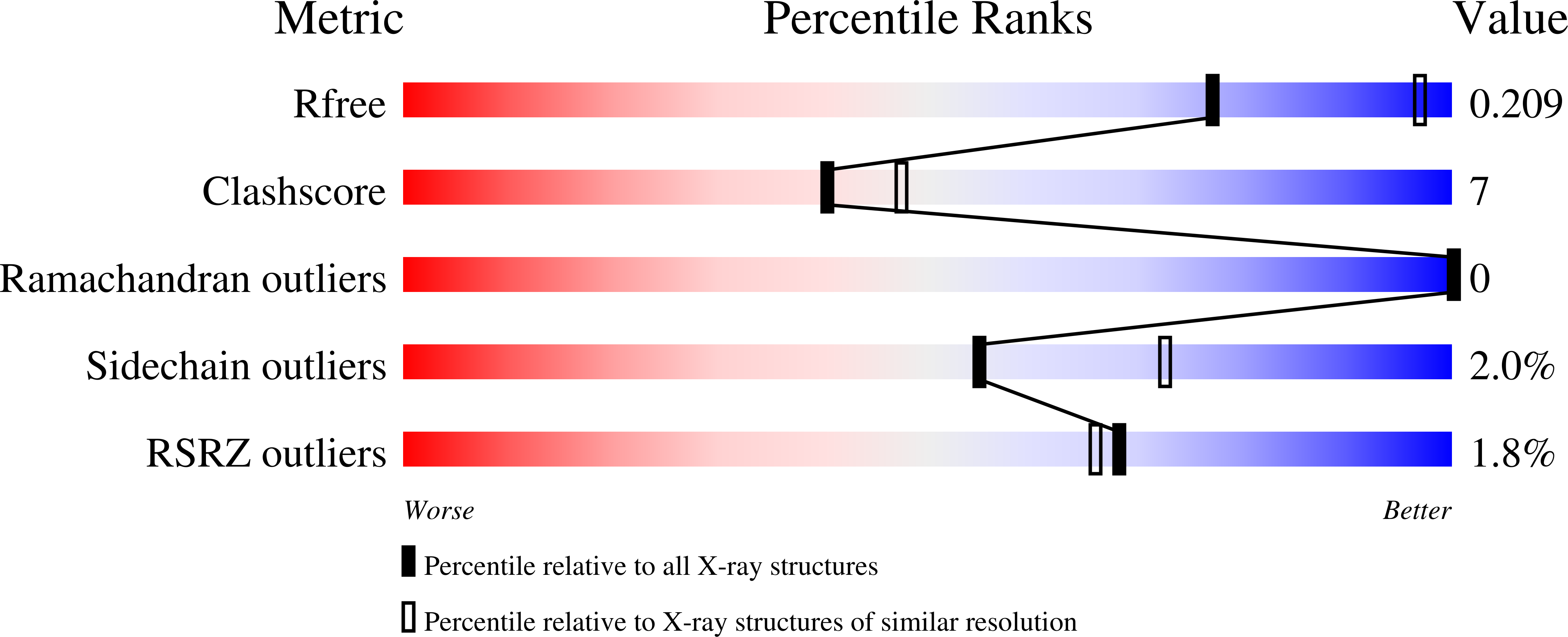
wwPDB Validation   3D Report Full Report


Ligand Structure Quality Assessment 


This is version 1.4 of the entry. See complete history


Literature

Structural and Biochemical Insights into the Inhibition of Human Acetylcholinesterase by G-Series Nerve Agents and Subsequent Reactivation by HI-6.

McGuire, J.R.Bester, S.M.Guelta, M.A.Cheung, J.Langley, C.Winemiller, M.D.Bae, S.Y.Funk, V.Myslinski, J.M.Pegan, S.D.Height, J.J.

(2021) Chem Res Toxicol 34: 804-816

  • DOI: https://doi.org/10.1021/acs.chemrestox.0c00406
  • Primary Citation of Related Structures:  
    6WUV, 6WUY, 6WUZ, 6WV1, 6WVC, 6WVO, 6WVP, 6WVQ

  • PubMed Abstract: 

    The recent use of organophosphate nerve agents in Syria, Malaysia, Russia, and the United Kingdom has reinforced the potential threat of their intentional release. These agents act through their ability to inhibit human acetylcholinesterase (hAChE; E.C. 3.1.1.7), an enzyme vital for survival. The toxicity of hAChE inhibition via G-series nerve agents has been demonstrated to vary widely depending on the G-agent used. To gain insight into this issue, the structures of hAChE inhibited by tabun, sarin, cyclosarin, soman, and GP were obtained along with the inhibition kinetics for these agents. Through this information, the role of hAChE active site plasticity in agent selectivity is revealed. With reports indicating that the efficacy of reactivators can vary based on the nerve agent inhibiting hAChE, human recombinatorially expressed hAChE was utilized to define these variations for HI-6 among various G-agents. To identify the structural underpinnings of this phenomenon, the structures of tabun, sarin, and soman-inhibited hAChE in complex with HI-6 were determined. This revealed how the presence of G-agent adducts impacts reactivator access and placement within the active site. These insights will contribute toward a path of next-generation reactivators and an improved understanding of the innate issues with the current reactivators.


  • Organizational Affiliation

    Department of Pharmaceutical and Biomedical Sciences, University of Georgia, Athens, Georgia 30602, United States.


Macromolecules
Find similar proteins by:  (by identity cutoff)  |  3D Structure
Entity ID: 1
MoleculeChains Sequence LengthOrganismDetailsImage
Acetylcholinesterase
A, B
542Homo sapiensMutation(s): 0 
Gene Names: ACHE
EC: 3.1.1.7
UniProt & NIH Common Fund Data Resources
Find proteins for P22303 (Homo sapiens)
Explore P22303 
Go to UniProtKB:  P22303
PHAROS:  P22303
GTEx:  ENSG00000087085 
Entity Groups  
Sequence Clusters30% Identity50% Identity70% Identity90% Identity95% Identity100% Identity
UniProt GroupP22303
Glycosylation
Glycosylation Sites: 3Go to GlyGen: P22303-1
Sequence Annotations
Expand
  • Reference Sequence
Oligosaccharides

Help

Entity ID: 2
MoleculeChains Length2D Diagram Glycosylation3D Interactions
2-acetamido-2-deoxy-beta-D-glucopyranose-(1-4)-[alpha-L-fucopyranose-(1-6)]2-acetamido-2-deoxy-beta-D-glucopyranose
C, D, E
3N-Glycosylation
Glycosylation Resources
GlyTouCan:  G21290RB
GlyCosmos:  G21290RB
GlyGen:  G21290RB
Experimental Data & Validation

Experimental Data

  • Method: X-RAY DIFFRACTION
  • Resolution: 2.63 Å
  • R-Value Free: 0.206 
  • R-Value Work: 0.170 
  • R-Value Observed: 0.172 
  • Space Group: P 31 2 1
Unit Cell:
Length ( Å )Angle ( ˚ )
a = 104.495α = 90
b = 104.495β = 90
c = 323.839γ = 120
Software Package:
Software NamePurpose
HKL-2000data reduction
HKL-2000data scaling
PHENIXrefinement
PDB_EXTRACTdata extraction
PHENIXphasing

Structure Validation

View Full Validation Report


Ligand Structure Quality Assessment 


Entry History & Funding Information

Deposition Data


Funding OrganizationLocationGrant Number
Defense Threat Reduction Agency (DTRA)United StatesCB#3889

Revision History  (Full details and data files)

  • Version 1.0: 2021-02-17
    Type: Initial release
  • Version 1.1: 2021-03-31
    Changes: Database references
  • Version 1.2: 2021-06-02
    Changes: Derived calculations
  • Version 1.3: 2023-10-18
    Changes: Data collection, Database references, Refinement description
  • Version 1.4: 2024-10-16
    Changes: Structure summary