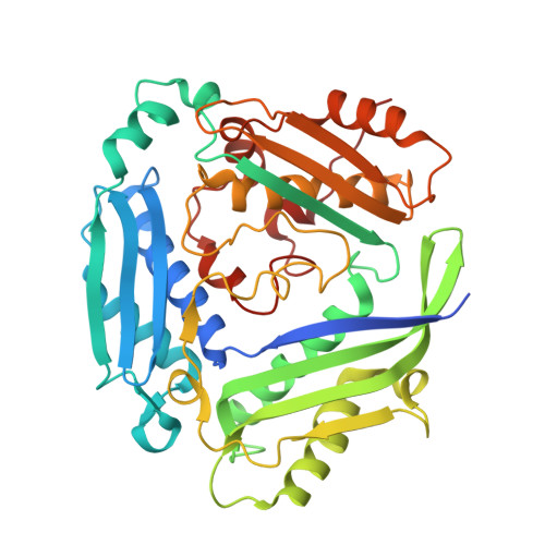
7BHW

Crystal structure of MAT2a bound to allosteric inhibitor (compound 29)


Experimental Data Snapshot

  • Method: X-RAY DIFFRACTION
  • Resolution: 1.15 Å
  • R-Value Free: 0.167 
  • R-Value Work: 0.155 
  • R-Value Observed: 0.156 

Starting Model: other
View more details

wwPDB Validation   3D Report Full Report


Ligand Structure Quality Assessment 


This is version 1.3 of the entry. See complete history


Literature

Fragment-Based Design of a Potent MAT2a Inhibitor and in Vivo Evaluation in an MTAP Null Xenograft Model.

De Fusco, C.Schimpl, M.Borjesson, U.Cheung, T.Collie, I.Evans, L.Narasimhan, P.Stubbs, C.Vazquez-Chantada, M.Wagner, D.J.Grondine, M.Sanders, M.G.Tentarelli, S.Underwood, E.Argyrou, A.Smith, J.M.Lynch, J.T.Chiarparin, E.Robb, G.Bagal, S.K.Scott, J.S.

(2021) J Med Chem 64: 6814-6826

  • DOI: https://doi.org/10.1021/acs.jmedchem.1c00067
  • Primary Citation of Related Structures:  
    7BHR, 7BHS, 7BHT, 7BHU, 7BHV, 7BHW, 7BHX

  • PubMed Abstract: 

    MAT2a is a methionine adenosyltransferase that synthesizes the essential metabolite S -adenosylmethionine (SAM) from methionine and ATP. Tumors bearing the co-deletion of p16 and MTAP genes have been shown to be sensitive to MAT2a inhibition, making it an attractive target for treatment of MTAP-deleted cancers. A fragment-based lead generation campaign identified weak but efficient hits binding in a known allosteric site. By use of structure-guided design and systematic SAR exploration, the hits were elaborated through a merging and growing strategy into an arylquinazolinone series of potent MAT2a inhibitors. The selected in vivo tool compound 28 reduced SAM-dependent methylation events in cells and inhibited proliferation of MTAP-null cells in vitro . In vivo studies showed that 28 was able to induce antitumor response in an MTAP knockout HCT116 xenograft model.


  • Organizational Affiliation

    Discovery Sciences, R&D, AstraZeneca, Cambridge CB4 0WG, United Kingdom.


Macromolecules
Find similar proteins by:  (by identity cutoff)  |  3D Structure
Entity ID: 1
MoleculeChains Sequence LengthOrganismDetailsImage
S-adenosylmethionine synthase isoform type-2397Homo sapiensMutation(s): 0 
Gene Names: MAT2AAMS2MATA2
EC: 2.5.1.6
UniProt & NIH Common Fund Data Resources
Find proteins for P31153 (Homo sapiens)
Explore P31153 
Go to UniProtKB:  P31153
PHAROS:  P31153
GTEx:  ENSG00000168906 
Entity Groups  
Sequence Clusters30% Identity50% Identity70% Identity90% Identity95% Identity100% Identity
UniProt GroupP31153
Sequence Annotations
Expand
  • Reference Sequence
Small Molecules
Ligands 2 Unique
IDChains Name / Formula / InChI Key2D Diagram3D Interactions
SAM
Query on SAM

Download Ideal Coordinates CCD File 
C [auth A]S-ADENOSYLMETHIONINE
C15 H22 N6 O5 S
MEFKEPWMEQBLKI-FCKMPRQPSA-N
TNW (Subject of Investigation/LOI)
Query on TNW

Download Ideal Coordinates CCD File 
B [auth A]7-chloranyl-4-(dimethylamino)-1-(3-methylphenyl)quinazolin-2-one
C17 H16 Cl N3 O
CQCXBTFANPRPTE-UHFFFAOYSA-N
Experimental Data & Validation

Experimental Data

  • Method: X-RAY DIFFRACTION
  • Resolution: 1.15 Å
  • R-Value Free: 0.167 
  • R-Value Work: 0.155 
  • R-Value Observed: 0.156 
  • Space Group: I 2 2 2
Unit Cell:
Length ( Å )Angle ( ˚ )
a = 68.58α = 90
b = 94.199β = 90
c = 117.404γ = 90
Software Package:
Software NamePurpose
XDSdata reduction
STARANISOdata scaling
AMoREphasing
BUSTERrefinement
PDB_EXTRACTdata extraction

Structure Validation

View Full Validation Report


Ligand Structure Quality Assessment 


Entry History 

Revision History  (Full details and data files)

  • Version 1.0: 2021-04-21
    Type: Initial release
  • Version 1.1: 2021-05-05
    Changes: Database references
  • Version 1.2: 2021-06-09
    Changes: Database references
  • Version 1.3: 2024-05-01
    Changes: Data collection, Database references, Refinement description