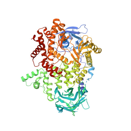
7BI9

PI3KC2a core in complex with PIK90


Experimental Data Snapshot

  • Method: X-RAY DIFFRACTION
  • Resolution: 2.65 Å
  • R-Value Free: 0.280 
  • R-Value Work: 0.233 
  • R-Value Observed: 0.235 

Starting Model: experimental
View more details

wwPDB Validation   3D Report Full Report


Ligand Structure Quality Assessment 


This is version 1.4 of the entry. See complete history


Literature

Structural basis of phosphatidylinositol 3-kinase C2 alpha function.

Lo, W.T.Zhang, Y.Vadas, O.Roske, Y.Gulluni, F.De Santis, M.C.Zagar, A.V.Stephanowitz, H.Hirsch, E.Liu, F.Daumke, O.Kudryashev, M.Haucke, V.

(2022) Nat Struct Mol Biol 29: 218-228

  • DOI: https://doi.org/10.1038/s41594-022-00730-w
  • Primary Citation of Related Structures:  
    7BI2, 7BI4, 7BI6, 7BI9

  • PubMed Abstract: 

    Phosphatidylinositol 3-kinase type 2α (PI3KC2α) is an essential member of the structurally unresolved class II PI3K family with crucial functions in lipid signaling, endocytosis, angiogenesis, viral replication, platelet formation and a role in mitosis. The molecular basis of these activities of PI3KC2α is poorly understood. Here, we report high-resolution crystal structures as well as a 4.4-Å cryogenic-electron microscopic (cryo-EM) structure of PI3KC2α in active and inactive conformations. We unravel a coincident mechanism of lipid-induced activation of PI3KC2α at membranes that involves large-scale repositioning of its Ras-binding and lipid-binding distal Phox-homology and C-C2 domains, and can serve as a model for the entire class II PI3K family. Moreover, we describe a PI3KC2α-specific helical bundle domain that underlies its scaffolding function at the mitotic spindle. Our results advance our understanding of PI3K biology and pave the way for the development of specific inhibitors of class II PI3K function with wide applications in biomedicine.


  • Organizational Affiliation

    Leibniz-Forschungsinstitut für Molekulare Pharmakologie (FMP), Berlin, Germany. lo@fmp-berlin.de.


Macromolecules
Find similar proteins by:  (by identity cutoff)  |  3D Structure
Entity ID: 1
MoleculeChains Sequence LengthOrganismDetailsImage
Phosphatidylinositol 4-phosphate 3-kinase C2 domain-containing subunit alpha,Phosphatidylinositol 4-phosphate 3-kinase C2 domain-containing subunit alpha910Mus musculusMutation(s): 3 
Gene Names: Pik3c2aCpk
EC: 2.7.1.154 (PDB Primary Data), 2.7.1.137 (UniProt), 2.7.1.153 (UniProt)
UniProt & NIH Common Fund Data Resources
Find proteins for Q61194 (Mus musculus)
Explore Q61194 
Go to UniProtKB:  Q61194
IMPC:  MGI:1203729
Entity Groups  
Sequence Clusters30% Identity50% Identity70% Identity90% Identity95% Identity100% Identity
UniProt GroupQ61194
Sequence Annotations
Expand
  • Reference Sequence
Experimental Data & Validation

Experimental Data

  • Method: X-RAY DIFFRACTION
  • Resolution: 2.65 Å
  • R-Value Free: 0.280 
  • R-Value Work: 0.233 
  • R-Value Observed: 0.235 
  • Space Group: P 21 21 21
Unit Cell:
Length ( Å )Angle ( ˚ )
a = 55.943α = 90
b = 135.397β = 90
c = 151.603γ = 90
Software Package:
Software NamePurpose
REFMACrefinement
PDB_EXTRACTdata extraction
XDSdata reduction
XSCALEdata scaling
PHASERphasing

Structure Validation

View Full Validation Report


Ligand Structure Quality Assessment 


Entry History 

Deposition Data

Revision History  (Full details and data files)

  • Version 1.0: 2022-03-09
    Type: Initial release
  • Version 1.1: 2022-03-23
    Changes: Database references
  • Version 1.2: 2022-03-30
    Changes: Database references
  • Version 1.3: 2022-04-27
    Changes: Database references
  • Version 1.4: 2024-01-31
    Changes: Data collection, Refinement description