7CYV

Crystal structure of FD20, a neutralizing single-chain variable fragment (scFv) in complex with SARS-CoV-2 Spike receptor-binding domain (RBD)


Experimental Data Snapshot

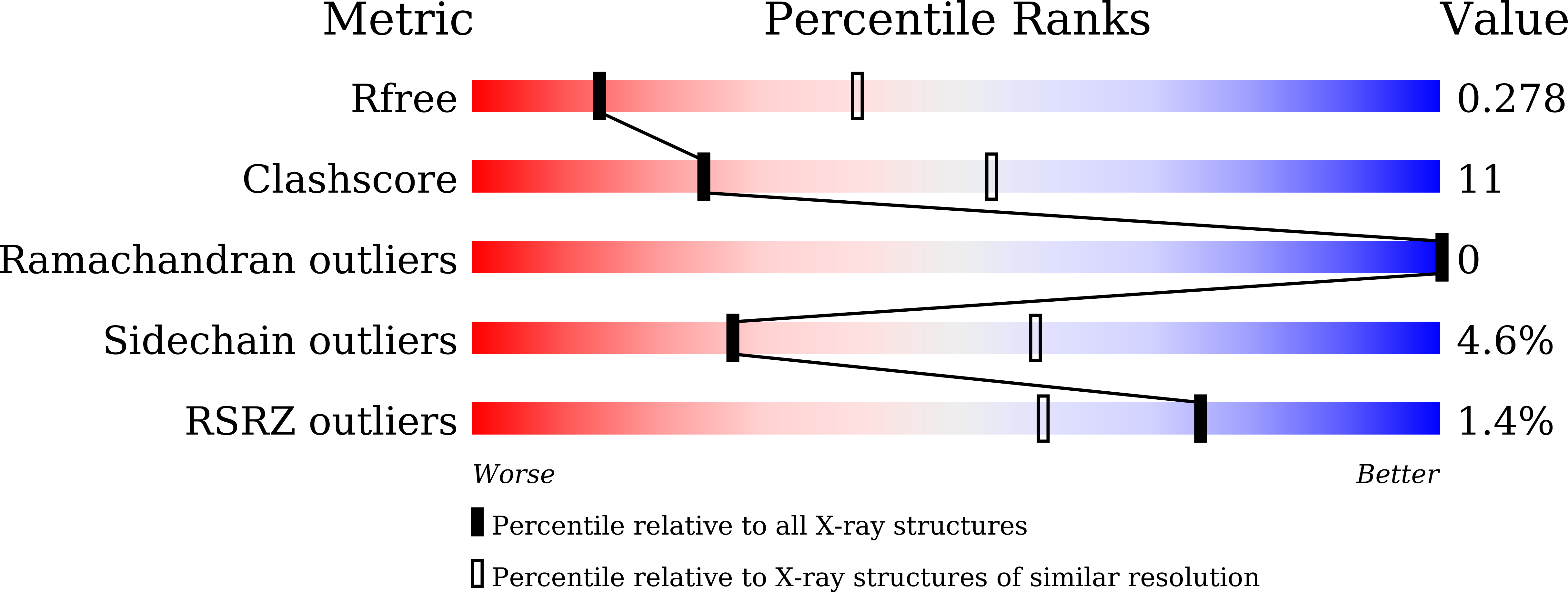
  • Method: X-RAY DIFFRACTION
  • Resolution: 3.13 Å
  • R-Value Free: 0.276 
  • R-Value Work: 0.252 
  • R-Value Observed: 0.253 

Starting Models: experimental
View more details

wwPDB Validation   3D Report Full Report


This is version 1.3 of the entry. See complete history


Literature

Uncovering a conserved vulnerability site in SARS-CoV-2 by a human antibody.

Li, T.Cai, H.Zhao, Y.Li, Y.Lai, Y.Yao, H.Liu, L.D.Sun, Z.van Vlissingen, M.F.Kuiken, T.GeurtsvanKessel, C.H.Zhang, N.Zhou, B.Lu, L.Gong, Y.Qin, W.Mondal, M.Duan, B.Xu, S.Richard, A.S.Raoul, H.Chen, J.Xu, C.Wu, L.Zhou, H.Huang, Z.Zhang, X.Li, J.Wang, Y.Bi, Y.Rockx, B.Chen, J.Meng, F.L.Lavillette, D.Li, D.

(2021) EMBO Mol Med 13: e14544-e14544

  • DOI: https://doi.org/10.15252/emmm.202114544
  • Primary Citation of Related Structures:  
    7CYV

  • PubMed Abstract: 

    An essential step for SARS-CoV-2 infection is the attachment to the host cell receptor by its Spike receptor-binding domain (RBD). Most of the existing RBD-targeting neutralizing antibodies block the receptor-binding motif (RBM), a mutable region with the potential to generate neutralization escape mutants. Here, we isolated and structurally characterized a non-RBM-targeting monoclonal antibody (FD20) from convalescent patients. FD20 engages the RBD at an epitope distal to the RBM with a K D of 5.6 nM, neutralizes SARS-CoV-2 including the current Variants of Concern such as B.1.1.7, B.1.351, P.1, and B.1.617.2 (Delta), displays modest cross-reactivity against SARS-CoV, and reduces viral replication in hamsters. The epitope coincides with a predicted "ideal" vulnerability site with high functional and structural constraints. Mutation of the residues of the conserved epitope variably affects FD20-binding but confers little or no resistance to neutralization. Finally, in vitro mode-of-action characterization and negative-stain electron microscopy suggest a neutralization mechanism by which FD20 destructs the Spike. Our results reveal a conserved vulnerability site in the SARS-CoV-2 Spike for the development of potential antiviral drugs.


  • Organizational Affiliation

    State Key Laboratory of Molecular Biology, State Key Laboratory of Cell Biology, CAS Center for Excellence in Molecular Cell Science, Shanghai Institute of Biochemistry and Cell Biology, Chinese Academy of Sciences (CAS), Shanghai, China.


Macromolecules
Find similar proteins by:  (by identity cutoff)  |  3D Structure
Entity ID: 1
MoleculeChains Sequence LengthOrganismDetailsImage
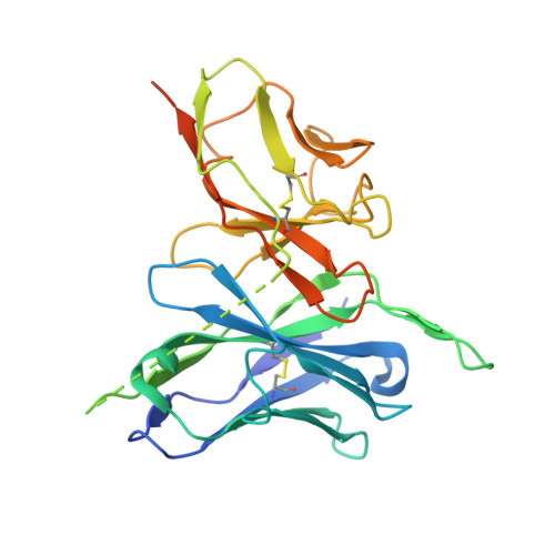
The heavy chain variable region of the scFv FD20,The light chain variable region of the scFv FD20A [auth H]281Homo sapiensMutation(s): 0 
Entity Groups  
Sequence Clusters30% Identity50% Identity70% Identity90% Identity95% Identity100% Identity
Sequence Annotations
Expand
  • Reference Sequence
Find similar proteins by:  (by identity cutoff)  |  3D Structure
Entity ID: 2
MoleculeChains Sequence LengthOrganismDetailsImage
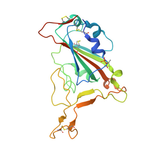
Spike protein S1213Severe acute respiratory syndrome coronavirus 2Mutation(s): 0 
Gene Names: S2
UniProt
Find proteins for P0DTC2 (Severe acute respiratory syndrome coronavirus 2)
Explore P0DTC2 
Go to UniProtKB:  P0DTC2
Entity Groups  
Sequence Clusters30% Identity50% Identity70% Identity90% Identity95% Identity100% Identity
UniProt GroupP0DTC2
Glycosylation
Glycosylation Sites: 1Go to GlyGen: P0DTC2-1
Sequence Annotations
Expand
  • Reference Sequence
Oligosaccharides

Help

Entity ID: 3
MoleculeChains Length2D Diagram Glycosylation3D Interactions
beta-D-mannopyranose-(1-4)-2-acetamido-2-deoxy-beta-D-glucopyranose-(1-4)-[alpha-L-fucopyranose-(1-3)][alpha-L-fucopyranose-(1-6)]2-acetamido-2-deoxy-beta-D-glucopyranoseC [auth A]5N-Glycosylation
Glycosylation Resources
GlyTouCan:  G92953ND
GlyCosmos:  G92953ND
Small Molecules
Modified Residues  1 Unique
IDChains TypeFormula2D DiagramParent
TPO
Query on TPO
B
L-PEPTIDE LINKINGC4 H10 N O6 PTHR
Experimental Data & Validation

Experimental Data

  • Method: X-RAY DIFFRACTION
  • Resolution: 3.13 Å
  • R-Value Free: 0.276 
  • R-Value Work: 0.252 
  • R-Value Observed: 0.253 
  • Space Group: C 1 2 1
Unit Cell:
Length ( Å )Angle ( ˚ )
a = 206.958α = 90
b = 57.925β = 100.433
c = 47.207γ = 90
Software Package:
Software NamePurpose
PHENIXrefinement
XDSdata reduction
XSCALEdata scaling
PHASERphasing

Structure Validation

View Full Validation Report


Entry History & Funding Information

Deposition Data


Funding OrganizationLocationGrant Number
National Science Foundation (NSF, China)China31870726

Revision History  (Full details and data files)

  • Version 1.0: 2021-09-15
    Type: Initial release
  • Version 1.1: 2022-03-30
    Changes: Database references
  • Version 1.2: 2023-11-29
    Changes: Data collection, Refinement description
  • Version 1.3: 2024-10-09
    Changes: Structure summary