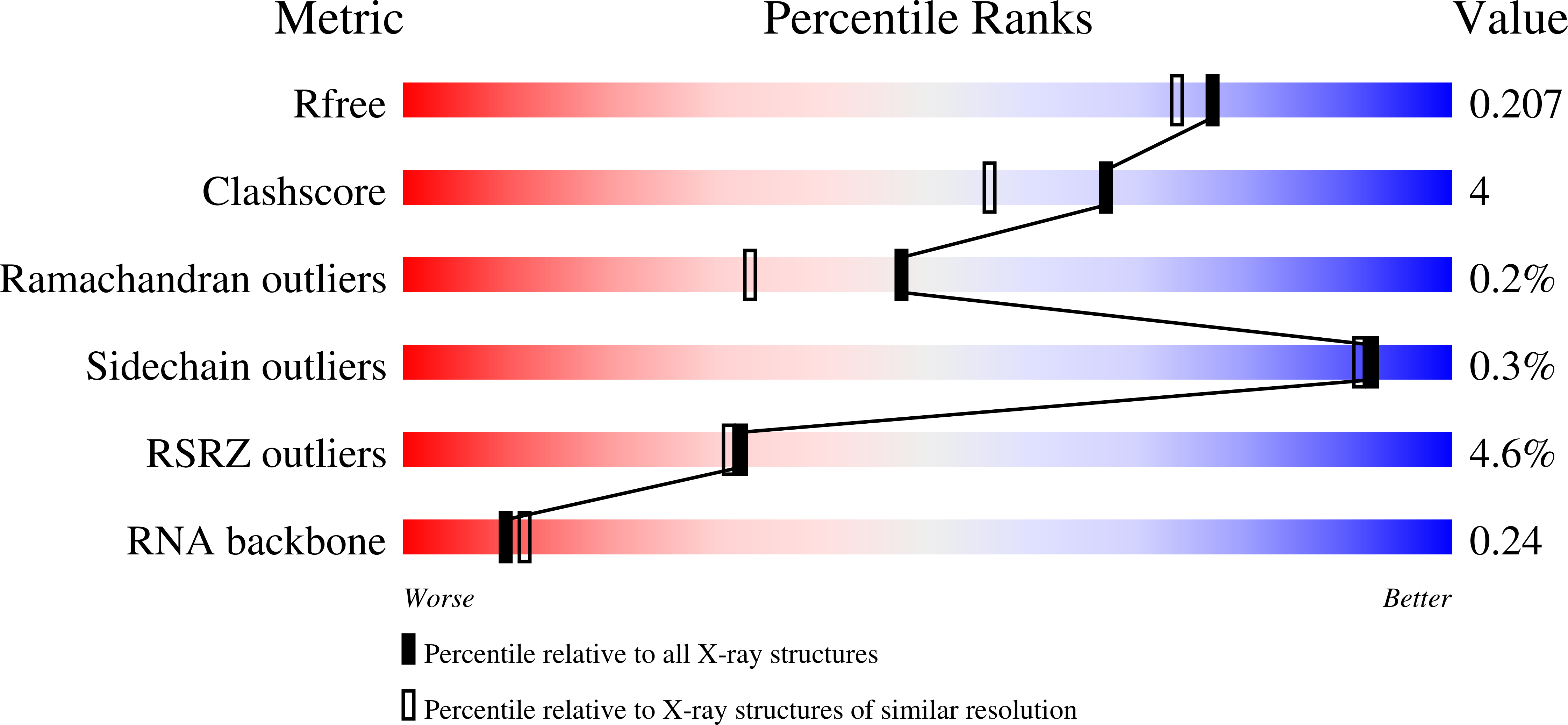
7MJY

MiaB in the complex with s-adenosyl-L-homocysteine and RNA


Experimental Data Snapshot

  • Method: X-RAY DIFFRACTION
  • Resolution: 1.86 Å
  • R-Value Free: 0.209 
  • R-Value Work: 0.174 
  • R-Value Observed: 0.176 

Starting Model: experimental
View more details

wwPDB Validation   3D Report Full Report


Ligand Structure Quality Assessment 


This is version 1.3 of the entry. See complete history


Literature

Structural basis for tRNA methylthiolation by the radical SAM enzyme MiaB.

Esakova, O.A.Grove, T.L.Yennawar, N.H.Arcinas, A.J.Wang, B.Krebs, C.Almo, S.C.Booker, S.J.

(2021) Nature 597: 566-570

  • DOI: https://doi.org/10.1038/s41586-021-03904-6
  • Primary Citation of Related Structures:  
    7MJV, 7MJW, 7MJX, 7MJY, 7MJZ

  • PubMed Abstract: 

    Numerous post-transcriptional modifications of transfer RNAs have vital roles in translation. The 2-methylthio-N 6 -isopentenyladenosine (ms 2 i 6 A) modification occurs at position 37 (A37) in transfer RNAs that contain adenine in position 36 of the anticodon, and serves to promote efficient A:U codon-anticodon base-pairing and to prevent unintended base pairing by near cognates, thus enhancing translational fidelity 1-4 . The ms 2 i 6 A modification is installed onto isopentenyladenosine (i 6 A) by MiaB, a radical S-adenosylmethionine (SAM) methylthiotransferase. As a radical SAM protein, MiaB contains one [Fe 4 S 4 ] RS cluster used in the reductive cleavage of SAM to form a 5'-deoxyadenosyl 5'-radical, which is responsible for removing the C 2 hydrogen of the substrate 5 . MiaB also contains an auxiliary [Fe 4 S 4 ] aux cluster, which has been implicated 6-9 in sulfur transfer to C 2 of i 6 A37. How this transfer takes place is largely unknown. Here we present several structures of MiaB from Bacteroides uniformis. These structures are consistent with a two-step mechanism, in which one molecule of SAM is first used to methylate a bridging µ-sulfido ion of the auxiliary cluster. In the second step, a second SAM molecule is cleaved to a 5'-deoxyadenosyl 5'-radical, which abstracts the C 2 hydrogen of the substrate but only after C 2 has undergone rehybridization from sp 2 to sp 3 . This work advances our understanding of how enzymes functionalize inert C-H bonds with sulfur.


  • Organizational Affiliation

    Department of Chemistry, The Pennsylvania State University, University Park, PA, USA. oae3@psu.edu.


Macromolecules

Find similar proteins by:  (by identity cutoff)  |  3D Structure
Entity ID: 1
MoleculeChains Sequence LengthOrganismDetailsImage
tRNA-2-methylthio-N(6)-dimethylallyladenosine synthase457Bacteroides uniformisMutation(s): 0 
Gene Names: 
EC: 2.8.4.3
UniProt
Find proteins for A0A174NUT3 (Bacteroides uniformis)
Explore A0A174NUT3 
Go to UniProtKB:  A0A174NUT3
Entity Groups  
Sequence Clusters30% Identity50% Identity70% Identity90% Identity95% Identity100% Identity
UniProt GroupA0A174NUT3
Sequence Annotations
Expand
  • Reference Sequence

Find similar nucleic acids by:  Sequence   |   3D Structure  

Entity ID: 2
MoleculeChains LengthOrganismImage
RNA (5'-R(P*UP*AP*UP*UP*C)-3')B [auth M]13Bacteroides uniformis
Sequence Annotations
Expand
  • Reference Sequence
Small Molecules
Ligands 6 Unique
IDChains Name / Formula / InChI Key2D Diagram3D Interactions
SAH (Subject of Investigation/LOI)
Query on SAH

Download Ideal Coordinates CCD File 
O [auth A]S-ADENOSYL-L-HOMOCYSTEINE
C14 H20 N6 O5 S
ZJUKTBDSGOFHSH-WFMPWKQPSA-N
SF4 (Subject of Investigation/LOI)
Query on SF4

Download Ideal Coordinates CCD File 
N [auth A]IRON/SULFUR CLUSTER
Fe4 S4
LJBDFODJNLIPKO-UHFFFAOYSA-N
F3S (Subject of Investigation/LOI)
Query on F3S

Download Ideal Coordinates CCD File 
M [auth A]FE3-S4 CLUSTER
Fe3 S4
FCXHZBQOKRZXKS-UHFFFAOYSA-N
PGE
Query on PGE

Download Ideal Coordinates CCD File 
C [auth A],
D [auth A],
E [auth A]
TRIETHYLENE GLYCOL
C6 H14 O4
ZIBGPFATKBEMQZ-UHFFFAOYSA-N
PEG
Query on PEG

Download Ideal Coordinates CCD File 
F [auth A]
G [auth A]
H [auth A]
I [auth A]
J [auth A]
F [auth A],
G [auth A],
H [auth A],
I [auth A],
J [auth A],
K [auth A],
L [auth A]
DI(HYDROXYETHYL)ETHER
C4 H10 O3
MTHSVFCYNBDYFN-UHFFFAOYSA-N
CL
Query on CL

Download Ideal Coordinates CCD File 
P [auth A]CHLORIDE ION
Cl
VEXZGXHMUGYJMC-UHFFFAOYSA-M
Experimental Data & Validation

Experimental Data

  • Method: X-RAY DIFFRACTION
  • Resolution: 1.86 Å
  • R-Value Free: 0.209 
  • R-Value Work: 0.174 
  • R-Value Observed: 0.176 
  • Space Group: P 21 21 21
Unit Cell:
Length ( Å )Angle ( ˚ )
a = 53.834α = 90
b = 79.982β = 90
c = 110.137γ = 90
Software Package:
Software NamePurpose
PHENIXrefinement
HKL-2000data scaling
PDB_EXTRACTdata extraction
HKL-2000data reduction
PHENIXphasing

Structure Validation

View Full Validation Report


Ligand Structure Quality Assessment 


Entry History & Funding Information

Deposition Data


Funding OrganizationLocationGrant Number
National Institutes of Health/National Institute of General Medical Sciences (NIH/NIGMS)United StatesNIH (GM-122595 to S.J.B)
National Science Foundation (NSF, United States)United StatesNSF (MCB-1716686 to S.J.B.)
Howard Hughes Medical Institute (HHMI)United StatesSquire J. Booker

Revision History  (Full details and data files)

  • Version 1.0: 2021-09-15
    Type: Initial release
  • Version 1.1: 2021-09-29
    Changes: Database references
  • Version 1.2: 2021-10-06
    Changes: Database references
  • Version 1.3: 2023-10-18
    Changes: Data collection, Refinement description