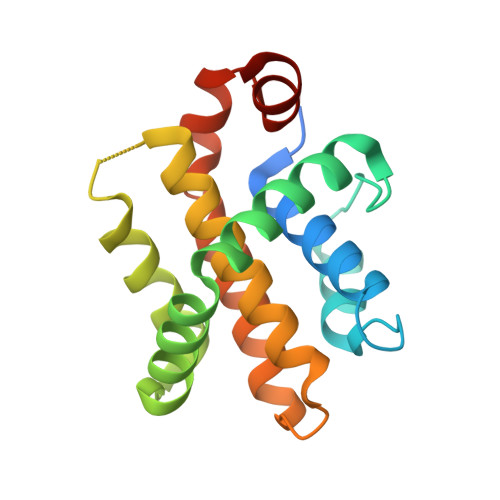
7P9W

Epstein-Barr virus encoded apoptosis regulator BHRF1 in complex with Puma BH3


Experimental Data Snapshot

  • Method: X-RAY DIFFRACTION
  • Resolution: 2.00 Å
  • R-Value Free: 0.257 
  • R-Value Work: 0.220 
  • R-Value Observed: 0.221 

Starting Model: experimental
View more details

wwPDB Validation   3D Report Full Report


This is version 1.2 of the entry. See complete history


Literature

Crystal Structures of Epstein-Barr Virus Bcl-2 Homolog BHRF1 Bound to Bid and Puma BH3 Motif Peptides.

Suraweera, C.D.Hinds, M.G.Kvansakul, M.

(2022) Viruses 14

  • DOI: https://doi.org/10.3390/v14102222
  • Primary Citation of Related Structures:  
    7P33, 7P9W

  • PubMed Abstract: 

    Apoptosis is a powerful defense mechanism used by multicellular organisms to counteract viral infection. In response to premature host cell suicide, viruses have evolved numerous countermeasures to ensure cell viability to optimize their replication by encoding proteins homologous in structure and function to cellular pro-survival Bcl-2 proteins. Epstein-Barr virus (EBV), a member of the Gammaherpesviridae , encodes the Bcl-2 homolog BHRF1, a potent inhibitor of Bcl-2-mediated apoptosis. BHRF1 acts by directly targeting Bid and Puma, two proapoptotic proteins of the Bcl-2 family. Here, we determined the crystal structures of BHRF1 bound to peptides spanning the Bcl-2 binding motifs (Bcl-2 homology 3 motif, BH3) of Bid and Puma. BHRF1 engages BH3 peptides using the canonical ligand-binding groove of its Bcl-2 fold and maintains a salt bridge between an Arg residue with a conserved Asp residue in the BH3 motif mimicking the canonical ionic interaction seen in host Bcl-2:BH3 motif complexes. Furthermore, both Bid and Puma utilize a fifth binding pocket in the canonical ligand binding groove of BHRF1 to provide an additional hydrophobic interaction distinct from the interactions previously seen with Bak and Bim. These findings provide a structural basis for EBV-mediated suppression of host cell apoptosis and reveal the flexibility of virus encoded Bcl-2 proteins in mimicking key interactions from the endogenous host signaling pathways.


  • Organizational Affiliation

    Department of Biochemistry & Chemistry, La Trobe Institute for Molecular Science, La Trobe University, Melbourne, VIC 3086, Australia.


Macromolecules
Find similar proteins by:  (by identity cutoff)  |  3D Structure
Entity ID: 1
MoleculeChains Sequence LengthOrganismDetailsImage
Apoptosis regulator BHRF1173Human herpesvirus 4 strain B95-8Mutation(s): 0 
Gene Names: BHRF1
UniProt
Find proteins for P03182 (Epstein-Barr virus (strain B95-8))
Explore P03182 
Go to UniProtKB:  P03182
Entity Groups  
Sequence Clusters30% Identity50% Identity70% Identity90% Identity95% Identity100% Identity
UniProt GroupP03182
Sequence Annotations
Expand
  • Reference Sequence
Find similar proteins by:  (by identity cutoff)  |  3D Structure
Entity ID: 2
MoleculeChains Sequence LengthOrganismDetailsImage
Bcl-2-binding component 3, isoforms 1/226Homo sapiensMutation(s): 0 
UniProt & NIH Common Fund Data Resources
Find proteins for Q9BXH1 (Homo sapiens)
Explore Q9BXH1 
Go to UniProtKB:  Q9BXH1
PHAROS:  Q9BXH1
GTEx:  ENSG00000105327 
Entity Groups  
Sequence Clusters30% Identity50% Identity70% Identity90% Identity95% Identity100% Identity
UniProt GroupQ9BXH1
Sequence Annotations
Expand
  • Reference Sequence
Small Molecules
Ligands 6 Unique
IDChains Name / Formula / InChI Key2D Diagram3D Interactions
PO4
Query on PO4

Download Ideal Coordinates CCD File 
C [auth A],
D [auth A],
E [auth A]
PHOSPHATE ION
O4 P
NBIIXXVUZAFLBC-UHFFFAOYSA-K
PDO
Query on PDO

Download Ideal Coordinates CCD File 
K [auth A],
M [auth A]
1,3-PROPANDIOL
C3 H8 O2
YPFDHNVEDLHUCE-UHFFFAOYSA-N
EDO
Query on EDO

Download Ideal Coordinates CCD File 
F [auth A]
G [auth A]
H [auth A]
I [auth A]
L [auth A]
F [auth A],
G [auth A],
H [auth A],
I [auth A],
L [auth A],
Y [auth B]
1,2-ETHANEDIOL
C2 H6 O2
LYCAIKOWRPUZTN-UHFFFAOYSA-N
CL
Query on CL

Download Ideal Coordinates CCD File 
S [auth A]
T [auth A]
U [auth A]
V [auth A]
W [auth A]
S [auth A],
T [auth A],
U [auth A],
V [auth A],
W [auth A],
X [auth A]
CHLORIDE ION
Cl
VEXZGXHMUGYJMC-UHFFFAOYSA-M
MG
Query on MG

Download Ideal Coordinates CCD File 
N [auth A],
O [auth A],
P [auth A],
Q [auth A],
R [auth A]
MAGNESIUM ION
Mg
JLVVSXFLKOJNIY-UHFFFAOYSA-N
NH4
Query on NH4

Download Ideal Coordinates CCD File 
J [auth A]AMMONIUM ION
H4 N
QGZKDVFQNNGYKY-UHFFFAOYSA-O
Experimental Data & Validation

Experimental Data

  • Method: X-RAY DIFFRACTION
  • Resolution: 2.00 Å
  • R-Value Free: 0.257 
  • R-Value Work: 0.220 
  • R-Value Observed: 0.221 
  • Space Group: P 32 2 1
Unit Cell:
Length ( Å )Angle ( ˚ )
a = 62.772α = 90
b = 62.772β = 90
c = 92.592γ = 120
Software Package:
Software NamePurpose
PHENIXrefinement
DIALSdata reduction
Aimlessdata scaling
PHASERphasing

Structure Validation

View Full Validation Report


Entry History & Funding Information

Deposition Data


Funding OrganizationLocationGrant Number
Australian Research Council (ARC)AustraliaFT130101349

Revision History  (Full details and data files)

  • Version 1.0: 2022-08-10
    Type: Initial release
  • Version 1.1: 2022-11-23
    Changes: Database references
  • Version 1.2: 2024-01-31
    Changes: Data collection, Refinement description