7U20 | pdb_00007u20

Crystal structure of human METTL1 and WDR4 complex


Experimental Data Snapshot

  • Method: X-RAY DIFFRACTION
  • Resolution: 3.10 Å
  • R-Value Free: 
    0.271 (Depositor), 0.270 (DCC) 
  • R-Value Work: 
    0.222 (Depositor), 0.230 (DCC) 
  • R-Value Observed: 
    0.224 (Depositor) 

Starting Models: experimental
View more details

wwPDB Validation   3D Report Full Report


Ligand Structure Quality Assessment 

Created with Raphaël 2.3.0Worse 01 BetterLigand structure goodness of fit to experimental dataBest fitted SO4Click on this verticalbar to view details

This is version 1.2 of the entry. See complete history


Literature

Structural basis of regulated m 7 G tRNA modification by METTL1-WDR4.

Li, J.Wang, L.Hahn, Q.Nowak, R.P.Viennet, T.Orellana, E.A.Roy Burman, S.S.Yue, H.Hunkeler, M.Fontana, P.Wu, H.Arthanari, H.Fischer, E.S.Gregory, R.I.

(2023) Nature 613: 391-397

  • DOI: https://doi.org/10.1038/s41586-022-05566-4
  • Primary Citation of Related Structures:  
    7U20, 8CTH, 8CTI

  • PubMed Abstract: 

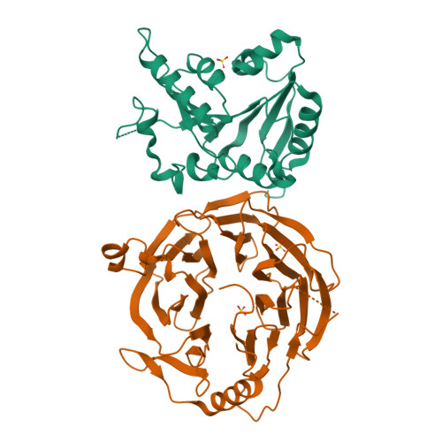
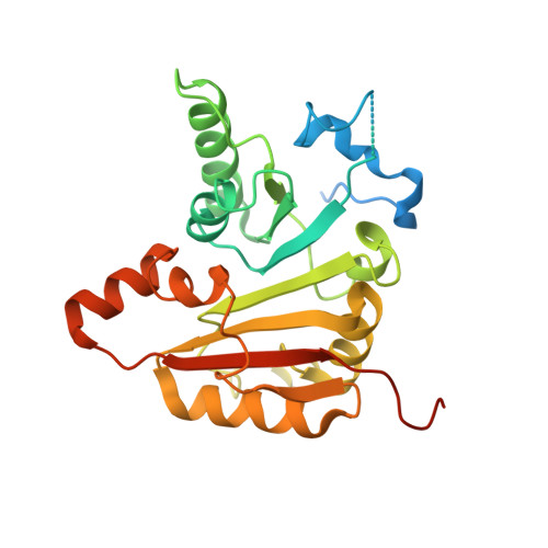
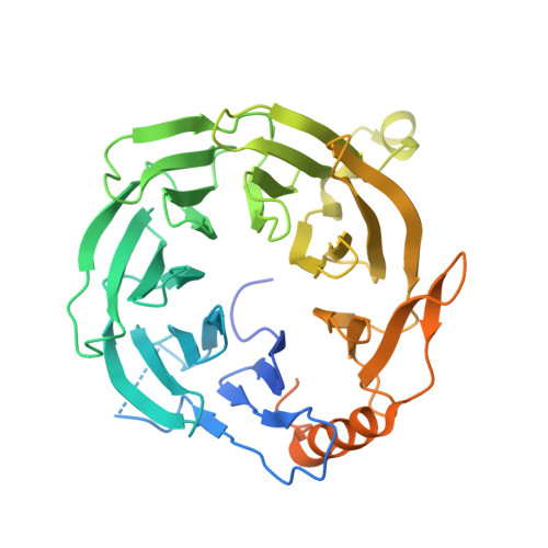
    Chemical modifications of RNA have key roles in many biological processes 1-3 . N 7 -methylguanosine (m 7 G) is required for integrity and stability of a large subset of tRNAs 4-7 . The methyltransferase 1-WD repeat-containing protein 4 (METTL1-WDR4) complex is the methyltransferase that modifies G46 in the variable loop of certain tRNAs, and its dysregulation drives tumorigenesis in numerous cancer types 8-14 . Mutations in WDR4 cause human developmental phenotypes including microcephaly 15-17 . How METTL1-WDR4 modifies tRNA substrates and is regulated remains elusive 18 . Here we show,  through structural, biochemical and cellular studies of human METTL1-WDR4, that WDR4 serves as a scaffold for METTL1 and the tRNA T-arm. Upon tRNA binding, the αC region of METTL1 transforms into a helix, which together with the α6 helix secures both ends of the tRNA variable loop. Unexpectedly, we find that the predicted disordered N-terminal region of METTL1 is part of the catalytic pocket and essential for methyltransferase activity. Furthermore, we reveal that S27 phosphorylation in the METTL1 N-terminal region inhibits methyltransferase activity by locally disrupting the catalytic centre. Our results provide a molecular understanding of tRNA substrate recognition and phosphorylation-mediated regulation of METTL1-WDR4, and reveal the presumed disordered N-terminal region of METTL1 as a nexus of methyltransferase activity.


  • Organizational Affiliation

    Stem Cell Program, Division of Hematology/Oncology, Boston Children's Hospital, Boston, MA, USA.


Macromolecules
Find similar proteins by:  (by identity cutoff)  |  3D Structure
Entity ID: 1
MoleculeChains Sequence LengthOrganismDetailsImage
tRNA (guanine-N(7)-)-methyltransferase276Homo sapiensMutation(s): 0 
Gene Names: METTL1C12orf1
EC: 2.1.1.33 (PDB Primary Data), 2.1.1 (PDB Primary Data)
UniProt & NIH Common Fund Data Resources
Find proteins for Q9UBP6 (Homo sapiens)
Explore Q9UBP6 
Go to UniProtKB:  Q9UBP6
PHAROS:  Q9UBP6
GTEx:  ENSG00000037897 
Entity Groups  
Sequence Clusters30% Identity50% Identity70% Identity90% Identity95% Identity100% Identity
UniProt GroupQ9UBP6
Sequence Annotations
Expand
  • Reference Sequence
Find similar proteins by:  (by identity cutoff)  |  3D Structure
Entity ID: 2
MoleculeChains Sequence LengthOrganismDetailsImage
tRNA (guanine-N(7)-)-methyltransferase non-catalytic subunit WDR4428Homo sapiensMutation(s): 0 
Gene Names: WDR4
UniProt & NIH Common Fund Data Resources
Find proteins for P57081 (Homo sapiens)
Explore P57081 
Go to UniProtKB:  P57081
PHAROS:  P57081
GTEx:  ENSG00000160193 
Entity Groups  
Sequence Clusters30% Identity50% Identity70% Identity90% Identity95% Identity100% Identity
UniProt GroupP57081
Sequence Annotations
Expand
  • Reference Sequence
Experimental Data & Validation

Experimental Data

  • Method: X-RAY DIFFRACTION
  • Resolution: 3.10 Å
  • R-Value Free:  0.271 (Depositor), 0.270 (DCC) 
  • R-Value Work:  0.222 (Depositor), 0.230 (DCC) 
  • R-Value Observed: 0.224 (Depositor) 
Space Group: C 2 2 21
Unit Cell:
Length ( Å )Angle ( ˚ )
a = 124.407α = 90
b = 194.574β = 90
c = 86.346γ = 90
Software Package:
Software NamePurpose
BUSTERrefinement
PHENIXrefinement
XDSdata reduction
Aimlessdata scaling
PHASERphasing

Structure Validation

View Full Validation Report


Ligand Structure Quality Assessment 

Created with Raphaël 2.3.0Worse 01 BetterLigand structure goodness of fit to experimental dataBest fitted SO4Click on this verticalbar to view details

Entry History & Funding Information

Deposition Data


Funding OrganizationLocationGrant Number
National Institutes of Health/National Cancer Institute (NIH/NCI)United States5R35CA232115
National Institutes of Health/National Cancer Institute (NIH/NCI)United StatesR01CA218278
National Institutes of Health/National Cancer Institute (NIH/NCI)United StatesR01CA214608

Revision History  (Full details and data files)

  • Version 1.0: 2022-12-07
    Type: Initial release
  • Version 1.1: 2023-02-15
    Changes: Database references
  • Version 1.2: 2023-10-25
    Changes: Data collection, Refinement description