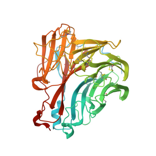
7U4G

Neuraminidase from influenza virus A/Shandong/9/1993(H3N2)


Experimental Data Snapshot

  • Method: X-RAY DIFFRACTION
  • Resolution: 1.65 Å
  • R-Value Free: 0.215 
  • R-Value Work: 0.188 
  • R-Value Observed: 0.189 

Starting Model: experimental
View more details

wwPDB Validation   3D Report Full Report


Ligand Structure Quality Assessment 


This is version 1.2 of the entry. See complete history


Literature

Prevalence and mechanisms of evolutionary contingency in human influenza H3N2 neuraminidase.

Lei, R.Tan, T.J.C.Hernandez Garcia, A.Wang, Y.Diefenbacher, M.Teo, C.Gopan, G.Tavakoli Dargani, Z.Teo, Q.W.Graham, C.S.Brooke, C.B.Nair, S.K.Wu, N.C.

(2022) Nat Commun 13: 6443-6443

  • DOI: https://doi.org/10.1038/s41467-022-34060-8
  • Primary Citation of Related Structures:  
    7U4E, 7U4F, 7U4G

  • PubMed Abstract: 

    Neuraminidase (NA) of human influenza H3N2 virus has evolved rapidly and been accumulating mutations for more than half-century. However, biophysical constraints that govern the evolutionary trajectories of NA remain largely elusive. Here, we show that among 70 natural mutations that are present in the NA of a recent human H3N2 strain, >10% are deleterious for an ancestral strain. By mapping the permissive mutations using combinatorial mutagenesis and next-generation sequencing, an extensive epistatic network is revealed. Biophysical and structural analyses further demonstrate that certain epistatic interactions can be explained by non-additive stability effect, which in turn modulates membrane trafficking and enzymatic activity of NA. Additionally, our results suggest that other biophysical mechanisms also contribute to epistasis in NA evolution. Overall, these findings not only provide mechanistic insights into the evolution of human influenza NA and elucidate its sequence-structure-function relationship, but also have important implications for the development of next-generation influenza vaccines.


  • Organizational Affiliation

    Department of Biochemistry, University of Illinois at Urbana-Champaign, Urbana, IL, 61801, USA.


Macromolecules
Find similar proteins by:  (by identity cutoff)  |  3D Structure
Entity ID: 1
MoleculeChains Sequence LengthOrganismDetailsImage
Neuraminidase469Influenza A virus (A/Shandong/9/1993(H3N2))Mutation(s): 0 
Gene Names: NA
EC: 3.2.1.18
UniProt
Find proteins for B8K0N8 (Influenza A virus)
Explore B8K0N8 
Go to UniProtKB:  B8K0N8
Entity Groups  
Sequence Clusters30% Identity50% Identity70% Identity90% Identity95% Identity100% Identity
UniProt GroupB8K0N8
Glycosylation
Glycosylation Sites: 3
Sequence Annotations
Expand
  • Reference Sequence
Oligosaccharides

Help

Entity ID: 2
MoleculeChains Length2D Diagram Glycosylation3D Interactions
alpha-D-mannopyranose-(1-2)-alpha-D-mannopyranose-(1-3)-[alpha-D-mannopyranose-(1-6)]beta-D-mannopyranose-(1-4)-2-acetamido-2-deoxy-beta-D-glucopyranose-(1-4)-2-acetamido-2-deoxy-beta-D-glucopyranose
B
6N-Glycosylation
Glycosylation Resources
GlyTouCan:  G56014GC
GlyCosmos:  G56014GC
GlyGen:  G56014GC
Entity ID: 3
MoleculeChains Length2D Diagram Glycosylation3D Interactions
alpha-D-mannopyranose-(1-4)-2-acetamido-2-deoxy-beta-D-glucopyranose
C
2N-Glycosylation
Glycosylation Resources
GlyTouCan:  G35893LO
GlyCosmos:  G35893LO
GlyGen:  G35893LO
Entity ID: 4
MoleculeChains Length2D Diagram Glycosylation3D Interactions
2-acetamido-2-deoxy-beta-D-glucopyranose-(1-4)-2-acetamido-2-deoxy-beta-D-glucopyranose
D
2N-Glycosylation
Glycosylation Resources
GlyTouCan:  G42666HT
GlyCosmos:  G42666HT
GlyGen:  G42666HT
Small Molecules
Ligands 2 Unique
IDChains Name / Formula / InChI Key2D Diagram3D Interactions
ZMR
Query on ZMR

Download Ideal Coordinates CCD File 
E [auth A]ZANAMIVIR
C12 H20 N4 O7
ARAIBEBZBOPLMB-UFGQHTETSA-N
CA
Query on CA

Download Ideal Coordinates CCD File 
F [auth A]CALCIUM ION
Ca
BHPQYMZQTOCNFJ-UHFFFAOYSA-N
Experimental Data & Validation

Experimental Data

  • Method: X-RAY DIFFRACTION
  • Resolution: 1.65 Å
  • R-Value Free: 0.215 
  • R-Value Work: 0.188 
  • R-Value Observed: 0.189 
  • Space Group: P 4 21 2
Unit Cell:
Length ( Å )Angle ( ˚ )
a = 107.954α = 90
b = 107.954β = 90
c = 78.619γ = 90
Software Package:
Software NamePurpose
REFMACrefinement
PDB_EXTRACTdata extraction
XDSdata reduction
autoPROCdata scaling
PHENIXphasing

Structure Validation

View Full Validation Report


Ligand Structure Quality Assessment 


Entry History & Funding Information

Deposition Data


Funding OrganizationLocationGrant Number
National Institutes of Health/National Institute Of Allergy and Infectious Diseases (NIH/NIAID)United StatesR00 AI139445
National Institutes of Health/National Institute Of Allergy and Infectious Diseases (NIH/NIAID)United StatesDP2 AT011966
National Institutes of Health/National Institute Of Allergy and Infectious Diseases (NIH/NIAID)United StatesR01 AI167910

Revision History  (Full details and data files)

  • Version 1.0: 2022-10-19
    Type: Initial release
  • Version 1.1: 2022-11-09
    Changes: Database references
  • Version 1.2: 2023-10-18
    Changes: Data collection, Refinement description