7WGT

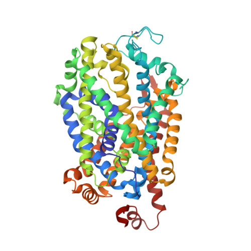
X-ray structure of thermostabilized Drosophila dopamine transporter with GABA transporter1-like substitutions in the binding site, in complex with NO711.


Experimental Data Snapshot

  • Method: X-RAY DIFFRACTION
  • Resolution: 2.75 Å
  • R-Value Free: 0.295 
  • R-Value Work: 0.248 
  • R-Value Observed: 0.250 

Starting Model: experimental
View more details

wwPDB Validation   3D Report Full Report


Ligand Structure Quality Assessment 


This is version 1.4 of the entry. See complete history

Re-refinement Note

This entry reflects an alternative modeling of the original data in: 7WGD


Literature

Structural insights into GABA transport inhibition using an engineered neurotransmitter transporter.

Joseph, D.Nayak, S.R.Penmatsa, A.

(2022) EMBO J 41: e110735-e110735

  • DOI: https://doi.org/10.15252/embj.2022110735
  • Primary Citation of Related Structures:  
    7WGD, 7WGT, 7WLW

  • PubMed Abstract: 

    γ-aminobutyric acid (GABA) is the major inhibitory neurotransmitter, and its levels in the synaptic space are controlled by the GABA transporter isoforms (GATs). GATs are structurally related to biogenic amine transporters but display interactions with distinct inhibitors used as anti-epileptics. In this study, we engineer the binding pocket of Drosophila melanogaster dopamine transporter to resemble GAT1 and determine high-resolution X-ray structures of the modified transporter in the substrate-free state and in complex with GAT1 inhibitors NO711 and SKF89976a that are analogs of tiagabine, a medication prescribed for the treatment of partial seizures. We observe that the primary binding site undergoes substantial shifts in subsite architecture in the modified transporter to accommodate the two GAT1 inhibitors. We also observe that SKF89976a additionally interacts at an allosteric site in the extracellular vestibule, yielding an occluded conformation. Interchanging SKF89976a interacting residue in the extracellular loop 4 between GAT1 and dDAT suggests a role for this motif in the selective control of neurotransmitter uptake. Our findings, therefore, provide vital insights into the organizational principles dictating GAT1 activity and inhibition.


  • Organizational Affiliation

    Molecular Biophysics Unit, Indian Institute of Science, Bangalore, India.


Macromolecules
Find similar proteins by:  (by identity cutoff)  |  3D Structure
Entity ID: 1
MoleculeChains Sequence LengthOrganismDetailsImage
Sodium-dependent dopamine transporter536Drosophila melanogasterMutation(s): 16 
Gene Names: DATfmnCG8380
Membrane Entity: Yes 
UniProt
Find proteins for Q7K4Y6 (Drosophila melanogaster)
Explore Q7K4Y6 
Go to UniProtKB:  Q7K4Y6
Entity Groups  
Sequence Clusters30% Identity50% Identity70% Identity90% Identity95% Identity100% Identity
UniProt GroupQ7K4Y6
Sequence Annotations
Expand
  • Reference Sequence
Find similar proteins by:  (by identity cutoff)  |  3D Structure
Entity ID: 2
MoleculeChains Sequence LengthOrganismDetailsImage
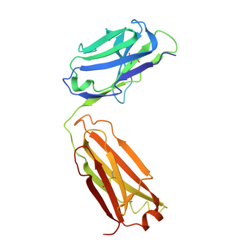
Antibody fragment (9D5) light chainB [auth L]214Mus musculusMutation(s): 0 
Entity Groups  
Sequence Clusters30% Identity50% Identity70% Identity90% Identity95% Identity100% Identity
Sequence Annotations
Expand
  • Reference Sequence
Find similar proteins by:  (by identity cutoff)  |  3D Structure
Entity ID: 3
MoleculeChains Sequence LengthOrganismDetailsImage
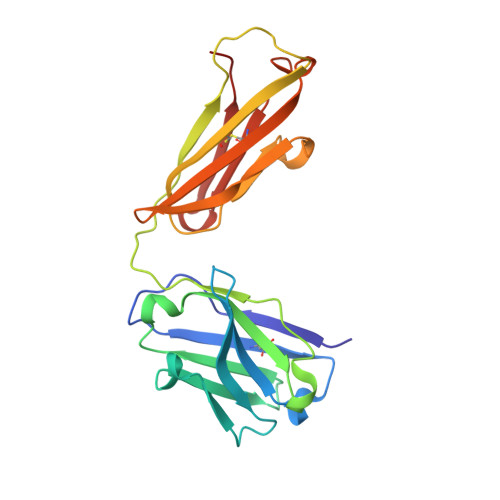
Antibody fragment (9D5) heavy chainC [auth H]219Mus musculusMutation(s): 0 
Entity Groups  
Sequence Clusters30% Identity50% Identity70% Identity90% Identity95% Identity100% Identity
Sequence Annotations
Expand
  • Reference Sequence
Small Molecules
Ligands 5 Unique
IDChains Name / Formula / InChI Key2D Diagram3D Interactions
Y01
Query on Y01

Download Ideal Coordinates CCD File 
I [auth A]CHOLESTEROL HEMISUCCINATE
C31 H50 O4
WLNARFZDISHUGS-MIXBDBMTSA-N
CLR
Query on CLR

Download Ideal Coordinates CCD File 
E [auth A]CHOLESTEROL
C27 H46 O
HVYWMOMLDIMFJA-DPAQBDIFSA-N
9BC (Subject of Investigation/LOI)
Query on 9BC

Download Ideal Coordinates CCD File 
D [auth A]1-(2-{[(diphenylmethylidene)amino]oxy}ethyl)-1,2,5,6-tetrahydropyridine-3-carboxylic acid
C21 H22 N2 O3
NGNALWDRPKNJGR-UHFFFAOYSA-N
CL
Query on CL

Download Ideal Coordinates CCD File 
H [auth A]CHLORIDE ION
Cl
VEXZGXHMUGYJMC-UHFFFAOYSA-M
NA
Query on NA

Download Ideal Coordinates CCD File 
F [auth A],
G [auth A]
SODIUM ION
Na
FKNQFGJONOIPTF-UHFFFAOYSA-N
Experimental Data & Validation

Experimental Data

  • Method: X-RAY DIFFRACTION
  • Resolution: 2.75 Å
  • R-Value Free: 0.295 
  • R-Value Work: 0.248 
  • R-Value Observed: 0.250 
  • Space Group: P 21 21 21
Unit Cell:
Length ( Å )Angle ( ˚ )
a = 98.007α = 90
b = 135.025β = 90
c = 162.559γ = 90
Software Package:
Software NamePurpose
PHENIXrefinement
XDSdata reduction
Aimlessdata scaling
PDB_EXTRACTdata extraction
PHASERphasing

Structure Validation

View Full Validation Report


Ligand Structure Quality Assessment 


Entry History & Funding Information

Deposition Data


Funding OrganizationLocationGrant Number
Department of Biotechnology (DBT, India)IndiaIA/I/15/2/502063

Revision History  (Full details and data files)

  • Version 1.0: 2022-06-29
    Type: Initial release
  • Version 1.1: 2022-07-20
    Changes: Database references
  • Version 1.2: 2022-08-17
    Changes: Database references
  • Version 1.3: 2023-11-29
    Changes: Data collection, Refinement description
  • Version 1.4: 2024-10-23
    Changes: Structure summary