7WPZ

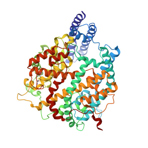
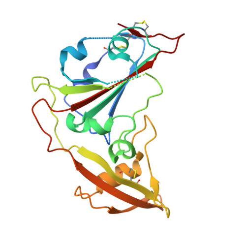
Structure of PDF-2180-COV RBD binding to Bat37 ACE2


Experimental Data Snapshot

  • Method: ELECTRON MICROSCOPY
  • Resolution: 3.80 Å
  • Aggregation State: PARTICLE 
  • Reconstruction Method: SINGLE PARTICLE 

wwPDB Validation   3D Report Full Report


This is version 1.5 of the entry. See complete history


Literature

Close relatives of MERS-CoV in bats use ACE2 as their functional receptors.

Xiong, Q.Cao, L.Ma, C.Tortorici, M.A.Liu, C.Si, J.Liu, P.Gu, M.Walls, A.C.Wang, C.Shi, L.Tong, F.Huang, M.Li, J.Zhao, C.Shen, C.Chen, Y.Zhao, H.Lan, K.Corti, D.Veesler, D.Wang, X.Yan, H.

(2022) Nature 612: 748-757

  • DOI: https://doi.org/10.1038/s41586-022-05513-3
  • Primary Citation of Related Structures:  
    7U6R, 7WPO, 7WPZ

  • PubMed Abstract: 

    Middle East respiratory syndrome coronavirus (MERS-CoV) and several bat coronaviruses use dipeptidyl peptidase-4 (DPP4) as an entry receptor 1-4 . However, the receptor for NeoCoV-the closest known MERS-CoV relative found in bats-remains unclear 5 . Here, using a pseudotype virus entry assay, we found that NeoCoV and its close relative, PDF-2180, can efficiently bind to and use specific bat angiotensin-converting enzyme 2 (ACE2) orthologues and, less favourably, human ACE2 as entry receptors through their receptor-binding domains (RBDs) on the spike (S) proteins. Cryo-electron microscopy analysis revealed an RBD-ACE2 binding interface involving protein-glycan interactions, distinct from those of other known ACE2-using coronaviruses. We identified residues 337-342 of human ACE2 as a molecular determinant restricting NeoCoV entry, whereas a NeoCoV S pseudotyped virus containing a T510F RBD mutation efficiently entered cells expressing human ACE2. Although polyclonal SARS-CoV-2 antibodies or MERS-CoV RBD-specific nanobodies did not cross-neutralize NeoCoV or PDF-2180, an ACE2-specific antibody and two broadly neutralizing betacoronavirus antibodies efficiently inhibited these two pseudotyped viruses. We describe MERS-CoV-related viruses that use ACE2 as an entry receptor, underscoring a promiscuity of receptor use and a potential zoonotic threat.


  • Organizational Affiliation

    State Key Laboratory of Virology, Institute for Vaccine Research and Modern Virology Research Center, College of Life Sciences, TaiKang Center for Life and Medical Sciences, Wuhan University, Wuhan, China.


Macromolecules
Find similar proteins by:  (by identity cutoff)  |  3D Structure
Entity ID: 1
MoleculeChains Sequence LengthOrganismDetailsImage
Angiotensin-converting enzyme595Pipistrellus pipistrellusMutation(s): 0 
Gene Names: mPipKuh1_000186
EC: 3.4
UniProt
Find proteins for A0A7J7V5I6 (Pipistrellus kuhlii)
Explore A0A7J7V5I6 
Go to UniProtKB:  A0A7J7V5I6
Entity Groups  
Sequence Clusters30% Identity50% Identity70% Identity90% Identity95% Identity100% Identity
UniProt GroupA0A7J7V5I6
Glycosylation
Glycosylation Sites: 7
Sequence Annotations
Expand
  • Reference Sequence
Find similar proteins by:  (by identity cutoff)  |  3D Structure
Entity ID: 2
MoleculeChains Sequence LengthOrganismDetailsImage
Spike glycoprotein204Bat coronavirusMutation(s): 0 
UniProt
Find proteins for A0A1W6ASU7 (Bat coronavirus)
Explore A0A1W6ASU7 
Go to UniProtKB:  A0A1W6ASU7
Entity Groups  
Sequence Clusters30% Identity50% Identity70% Identity90% Identity95% Identity100% Identity
UniProt GroupA0A1W6ASU7
Glycosylation
Glycosylation Sites: 1
Sequence Annotations
Expand
  • Reference Sequence
Oligosaccharides

Help

Entity ID: 3
MoleculeChains Length2D Diagram Glycosylation3D Interactions
2-acetamido-2-deoxy-beta-D-glucopyranose-(1-4)-2-acetamido-2-deoxy-beta-D-glucopyranoseC [auth E],
D [auth J],
F [auth D]
2N-Glycosylation
Glycosylation Resources
GlyTouCan:  G42666HT
GlyCosmos:  G42666HT
GlyGen:  G42666HT
Entity ID: 4
MoleculeChains Length2D Diagram Glycosylation3D Interactions
alpha-D-mannopyranose-(1-3)-[alpha-D-mannopyranose-(1-6)]beta-D-mannopyranose-(1-4)-2-acetamido-2-deoxy-beta-D-glucopyranose-(1-4)-2-acetamido-2-deoxy-beta-D-glucopyranoseE [auth C]5N-Glycosylation
Glycosylation Resources
GlyTouCan:  G22768VO
GlyCosmos:  G22768VO
GlyGen:  G22768VO
Experimental Data & Validation

Experimental Data

  • Method: ELECTRON MICROSCOPY
  • Resolution: 3.80 Å
  • Aggregation State: PARTICLE 
  • Reconstruction Method: SINGLE PARTICLE 

Structure Validation

View Full Validation Report


Entry History & Funding Information

Deposition Data


Funding OrganizationLocationGrant Number
Not funded--

Revision History  (Full details and data files)

  • Version 1.0: 2022-11-30
    Type: Initial release
  • Version 1.1: 2022-12-21
    Changes: Database references
  • Version 1.2: 2022-12-28
    Changes: Database references
  • Version 1.3: 2023-01-04
    Changes: Database references
  • Version 1.4: 2023-03-15
    Changes: Structure summary
  • Version 1.5: 2024-10-16
    Changes: Data collection, Structure summary