8AY0

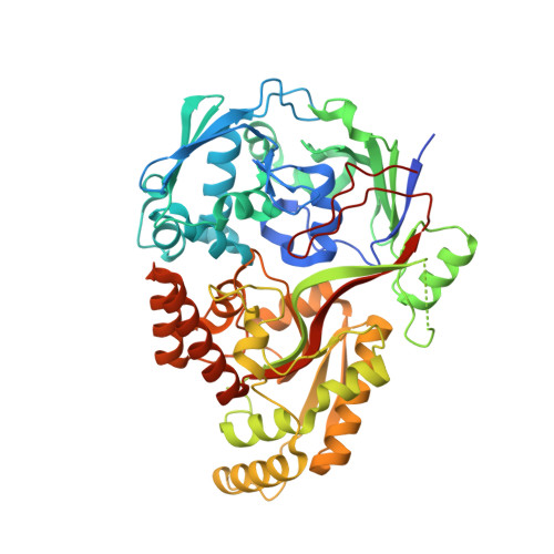
Crystal Structure of the peptide binding protein DppE from Bacillus subtilis in complex with murein tripeptide


Experimental Data Snapshot

  • Method: X-RAY DIFFRACTION
  • Resolution: 1.51 Å
  • R-Value Free: 0.221 
  • R-Value Work: 0.184 

Starting Model: experimental
View more details

wwPDB Validation   3D Report Full Report


Ligand Structure Quality Assessment 


This is version 1.1 of the entry. See complete history


Literature

Peptide transport in Bacillus subtilis - structure and specificity in the extracellular solute binding proteins OppA and DppE.

Hughes, A.M.Darby, J.F.Dodson, E.J.Wilson, S.J.Turkenburg, J.P.Thomas, G.H.Wilkinson, A.J.

(2022) Microbiology (Reading) 168

  • DOI: https://doi.org/10.1099/mic.0.001274
  • Primary Citation of Related Structures:  
    8ARE, 8ARN, 8AY0, 8AZB

  • PubMed Abstract: 

    Peptide transporters play important nutritional and cell signalling roles in Bacillus subtilis, which are pronounced during stationary phase adaptations and development. Three high-affinity ATP-binding cassette (ABC) family transporters are involved in peptide uptake - the oligopeptide permease (Opp), another peptide permease (App) and a less well-characterized dipeptide permease (Dpp). Here we report crystal structures of the extracellular substrate binding proteins, OppA and DppE, which serve the Opp and Dpp systems, respectively. The structure of OppA was determined in complex with endogenous peptides, modelled as Ser-Asn-Ser-Ser, and with the sporulation-promoting peptide Ser-Arg-Asn-Val-Thr, which bind with K d values of 0.4 and 2 µM, respectively, as measured by isothermal titration calorimetry. Differential scanning fluorescence experiments with a wider panel of ligands showed that OppA has highest affinity for tetra- and penta-peptides. The structure of DppE revealed the unexpected presence of a murein tripeptide (MTP) ligand, l-Ala-d-Glu- meso -DAP, in the peptide binding groove. The mode of MTP binding in DppE is different to that observed in the murein peptide binding protein, MppA, from Escherichia coli , suggesting independent evolution of these proteins from an OppA-like precursor. The presence of MTP in DppE points to a role for Dpp in the uptake and recycling of cell wall peptides, a conclusion that is supported by analysis of the genomic context of dpp , which revealed adjacent genes encoding enzymes involved in muropeptide catabolism in a gene organization that is widely conserved in Firmicutes .


  • Organizational Affiliation

    Structural Biology Laboratory, Department of Chemistry, University of York, York YO10 5DD, UK.


Macromolecules
Find similar proteins by:  (by identity cutoff)  |  3D Structure
Entity ID: 1
MoleculeChains Sequence LengthOrganismDetailsImage
Dipeptide-binding protein DppE
A, B, C
521Bacillus subtilisMutation(s): 0 
Gene Names: dppEdciAEBSU12960
UniProt
Find proteins for P26906 (Bacillus subtilis (strain 168))
Explore P26906 
Go to UniProtKB:  P26906
Entity Groups  
Sequence Clusters30% Identity50% Identity70% Identity90% Identity95% Identity100% Identity
UniProt GroupP26906
Sequence Annotations
Expand
  • Reference Sequence
Small Molecules
Ligands 3 Unique
IDChains Name / Formula / InChI Key2D Diagram3D Interactions
MHI (Subject of Investigation/LOI)
Query on MHI

Download Ideal Coordinates CCD File 
BA [auth C],
D [auth A],
S [auth B]
L-ALA-GAMMA-D-GLU-MESO-DIAMINOPIMELIC ACID
C15 H26 N4 O8
FMNCPUGORYYCEM-QCLAVDOMSA-N
EDO
Query on EDO

Download Ideal Coordinates CCD File 
AA [auth B]
CA [auth C]
DA [auth C]
EA [auth C]
F [auth A]
AA [auth B],
CA [auth C],
DA [auth C],
EA [auth C],
F [auth A],
FA [auth C],
G [auth A],
GA [auth C],
H [auth A],
I [auth A],
J [auth A],
K [auth A],
L [auth A],
M [auth A],
N [auth A],
O [auth A],
P [auth A],
Q [auth A],
R [auth A],
W [auth B],
X [auth B],
Y [auth B],
Z [auth B]
1,2-ETHANEDIOL
C2 H6 O2
LYCAIKOWRPUZTN-UHFFFAOYSA-N
MG
Query on MG

Download Ideal Coordinates CCD File 
E [auth A],
T [auth B],
U [auth B],
V [auth B]
MAGNESIUM ION
Mg
JLVVSXFLKOJNIY-UHFFFAOYSA-N
Experimental Data & Validation

Experimental Data

  • Method: X-RAY DIFFRACTION
  • Resolution: 1.51 Å
  • R-Value Free: 0.221 
  • R-Value Work: 0.184 
  • Space Group: P 1
Unit Cell:
Length ( Å )Angle ( ˚ )
a = 60.855α = 78.088
b = 61.31β = 82.672
c = 124.073γ = 61.597
Software Package:
Software NamePurpose
REFMACrefinement
REFMACrefinement
autoPROCdata processing
XDSdata reduction
Aimlessdata scaling
MOLREPphasing

Structure Validation

View Full Validation Report


Ligand Structure Quality Assessment 


Entry History & Funding Information

Deposition Data


Funding OrganizationLocationGrant Number
Biotechnology and Biological Sciences Research Council (BBSRC)United Kingdom--

Revision History  (Full details and data files)

  • Version 1.0: 2023-02-22
    Type: Initial release
  • Version 1.1: 2024-02-07
    Changes: Data collection, Refinement description