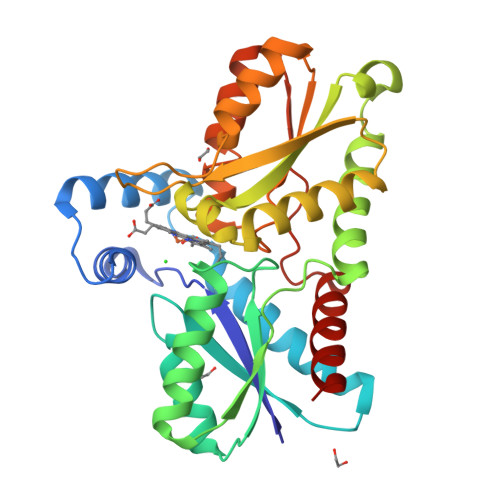
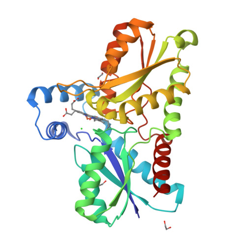
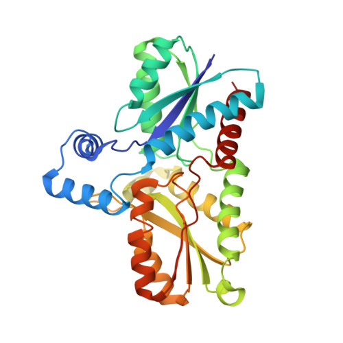
8BBV

Coproporphyrin III - LmCpfC complex soaked 2min with Fe2+


Experimental Data Snapshot

  • Method: X-RAY DIFFRACTION
  • Resolution: 2.19 Å
  • R-Value Free: 0.242 
  • R-Value Work: 0.192 
  • R-Value Observed: 0.195 

Starting Model: experimental
View more details

wwPDB Validation   3D Report Full Report


Ligand Structure Quality Assessment 


This is version 2.2 of the entry. See complete history


Literature

Iron insertion into coproporphyrin III-ferrochelatase complex: Evidence for an intermediate distorted catalytic species.

Gabler, T.Dali, A.Sebastiani, F.Furtmuller, P.G.Becucci, M.Hofbauer, S.Smulevich, G.

(2023) Protein Sci 32: e4788-e4788

  • DOI: https://doi.org/10.1002/pro.4788
  • Primary Citation of Related Structures:  
    8BBV, 8OFL, 8OMM

  • PubMed Abstract: 

    Understanding the reaction mechanism of enzymes at the molecular level is generally a difficult task, since many parameters affect the turnover. Often, due to high reactivity and formation of transient species or intermediates, detailed information on enzymatic catalysis is obtained by means of model substrates. Whenever possible, it is essential to confirm a reaction mechanism based on substrate analogues or model systems by using the physiological substrates. Here we disclose the ferrous iron incorporation mechanism, in solution, and in crystallo, by the coproporphyrin III-coproporphyrin ferrochelatase complex from the firmicute, pathogen, and antibiotic resistant, Listeria monocytogenes. Coproporphyrin ferrochelatase plays an important physiological role as the metalation represents the penultimate reaction step in the prokaryotic coproporphyrin-dependent heme biosynthetic pathway, yielding coproheme (ferric coproporphyrin III). By following the metal titration with resonance Raman spectroscopy and x-ray crystallography, we prove that upon metalation the saddling distortion becomes predominant both in the crystal and in solution. This is a consequence of the readjustment of hydrogen bond interactions of the propionates with the protein scaffold during the enzymatic catalysis. Once the propionates have established the interactions typical of the coproheme complex, the distortion slowly decreases, to reach the almost planar final product.


  • Organizational Affiliation

    Department of Chemistry, Institute of Biochemistry, University of Natural Resources and Life Sciences, Vienna, Austria.


Macromolecules
Find similar proteins by:  (by identity cutoff)  |  3D Structure
Entity ID: 1
MoleculeChains Sequence LengthOrganismDetailsImage
Coproporphyrin III ferrochelatase311Listeria monocytogenesMutation(s): 0 
Gene Names: cpfChemHlmo2211
EC: 4.99.1.9
UniProt
Find proteins for Q8Y565 (Listeria monocytogenes serovar 1/2a (strain ATCC BAA-679 / EGD-e))
Explore Q8Y565 
Go to UniProtKB:  Q8Y565
Entity Groups  
Sequence Clusters30% Identity50% Identity70% Identity90% Identity95% Identity100% Identity
UniProt GroupQ8Y565
Sequence Annotations
Expand
  • Reference Sequence
Small Molecules
Ligands 5 Unique
IDChains Name / Formula / InChI Key2D Diagram3D Interactions
HT9 (Subject of Investigation/LOI)
Query on HT9

Download Ideal Coordinates CCD File 
E [auth A]coproporphyrin III
C36 H38 N4 O8
XNBNKCLBGTWWSD-UJJXFSCMSA-N
GOL
Query on GOL

Download Ideal Coordinates CCD File 
B [auth A]GLYCEROL
C3 H8 O3
PEDCQBHIVMGVHV-UHFFFAOYSA-N
EDO
Query on EDO

Download Ideal Coordinates CCD File 
C [auth A],
D [auth A]
1,2-ETHANEDIOL
C2 H6 O2
LYCAIKOWRPUZTN-UHFFFAOYSA-N
FE2
Query on FE2

Download Ideal Coordinates CCD File 
F [auth A]FE (II) ION
Fe
CWYNVVGOOAEACU-UHFFFAOYSA-N
CL
Query on CL

Download Ideal Coordinates CCD File 
G [auth A]CHLORIDE ION
Cl
VEXZGXHMUGYJMC-UHFFFAOYSA-M
Experimental Data & Validation

Experimental Data

  • Method: X-RAY DIFFRACTION
  • Resolution: 2.19 Å
  • R-Value Free: 0.242 
  • R-Value Work: 0.192 
  • R-Value Observed: 0.195 
  • Space Group: P 1 21 1
Unit Cell:
Length ( Å )Angle ( ˚ )
a = 36.615α = 90
b = 66.919β = 103.218
c = 61.802γ = 90
Software Package:
Software NamePurpose
PHENIXrefinement
PHENIXrefinement
XDSdata reduction
XDSdata scaling
PHASERphasing

Structure Validation

View Full Validation Report


Ligand Structure Quality Assessment 


Entry History & Funding Information

Deposition Data


Funding OrganizationLocationGrant Number
Austrian Science FundAustriaP33544

Revision History  (Full details and data files)

  • Version 1.0: 2023-01-11
    Type: Initial release
  • Version 2.0: 2023-04-12
    Changes: Advisory, Atomic model, Data collection, Derived calculations, Non-polymer description, Structure summary
  • Version 2.1: 2023-12-06
    Changes: Data collection, Database references
  • Version 2.2: 2024-02-07
    Changes: Refinement description