Structure of Leishmania donovani 6-Phosphogluconate Dehydrogenase and Inhibition by Phosphine Gold(I) Complexes: A Potential Approach to Leishmaniasis Treatment.
Berneburg, I., Stumpf, M., Velten, A.S., Rahlfs, S., Przyborski, J., Becker, K., Fritz-Wolf, K.(2023) Int J Mol Sci 24
- PubMed: 37239962
- DOI: https://doi.org/10.3390/ijms24108615
- Primary Citation of Related Structures:
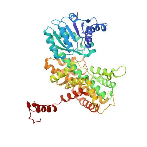
8C79 - PubMed Abstract:
As unicellular parasites are highly dependent on NADPH as a source for reducing equivalents, the main NADPH-producing enzymes glucose 6-phosphate dehydrogenase (G6PD) and 6-phosphogluconate dehydrogenase (6PGD) of the pentose phosphate pathway are considered promising antitrypanosomatid drug targets. Here we present the biochemical characterization and crystal structure of Leishmania donovani 6PGD ( Ld 6PGD) in complex with NADP(H). Most interestingly, a previously unknown conformation of NADPH is visible in this structure. In addition, we identified auranofin and other gold(I)-containing compounds as efficient Ld 6PGD inhibitors, although it has so far been assumed that trypanothione reductase is the sole target of auranofin in Kinetoplastida . Interestingly, 6PGD from Plasmodium falciparum is also inhibited at lower micromolar concentrations, whereas human 6PGD is not. Mode-of-inhibition studies indicate that auranofin competes with 6PG for its binding site followed by a rapid irreversible inhibition. By analogy with other enzymes, this suggests that the gold moiety is responsible for the observed inhibition. Taken together, we identified gold(I)-containing compounds as an interesting class of inhibitors against 6PGDs from Leishmania and possibly from other protozoan parasites. Together with the three-dimensional crystal structure, this provides a valid basis for further drug discovery approaches.
Organizational Affiliation:
Biochemistry and Molecular Biology, Interdisciplinary Research Center, Justus Liebig University, 35392 Giessen, Germany.