8DHT

Crystal structure of a typeIII Rubisco

  • Classification: LYASE
  • Organism(s): Archaeoglobus fulgidus
  • Expression System: Escherichia coli
  • Mutation(s): No 

  • Deposited: 2022-06-28 Released: 2022-11-30 
  • Deposition Author(s): Qingqiu, H.
  • Funding Organization(s): National Institutes of Health/National Institute of General Medical Sciences (NIH/NIGMS)

Experimental Data Snapshot

  • Method: X-RAY DIFFRACTION
  • Resolution: 1.70 Å
  • R-Value Free: 0.177 
  • R-Value Work: 0.144 
  • R-Value Observed: 0.144 

wwPDB Validation   3D Report Full Report


Ligand Structure Quality Assessment 


This is version 1.3 of the entry. See complete history


Literature

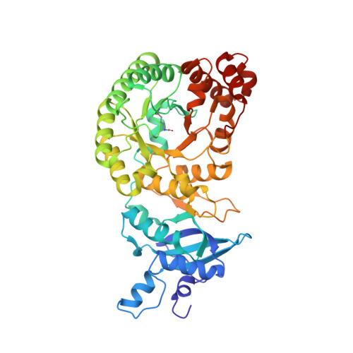
Crystal structure of a type III Rubisco in complex with its product 3-phosphoglycerate.

Huang, Q.Szebenyi, D.M.E.

(2023) Proteins 91: 330-337

  • DOI: https://doi.org/10.1002/prot.26431
  • Primary Citation of Related Structures:  
    8DHT

  • PubMed Abstract: 

    The crystal structure of the complex of ribulose-1,5-bisphosphate carboxylase/oxygenase (Rubisco; EC 4.1.1.39) from Archaeoglobus fulgidus (afRubisco) with its products 3PGAs has been determined to a resolution of 1.7 Å and is of the closed form. Type III Rubiscos such as afRubisco have 18 out of the 19 essential amino acid residues of canonical Rubisco; the 19th is Tyr rather than Phe. Superposition with the structure of a complex of the similar tkRubisco with the six-carbon intermediate analog 2CABP shows the same conformation of the 19 residues except for Glu46 and Thr51. Glu46 adopts a unique conformation different from that in other Rubiscos and makes two H-bonds with the ligand 3PGA. Similar to other closed state Rubiscos, the backbone of Thr51 is rotated and the side chain makes an H-bond with the ligand 3PGA. Two product 3PGA molecules are bound at the active site, overlapping well with the 2CABP of tkRubisco/2CABP. The positions of the P1 and P2 phosphate groups differ by 0.4 and 0.53 Å, respectively, between 2CABP and the two 3PGAs. This afRubisco/3PGA complex mimics an intermediate stage of the carboxylation reaction which occurs after the production of the two 3PGA products but before the reopening of the active site. The stability of this complex suggests that the Rubisco active site will not reopen before both 3PGA products are formed.


  • Organizational Affiliation

    Cornell High Energy Synchrotron Source (CHESS), Cornell University, Ithaca, New York, USA.


Macromolecules
Find similar proteins by:  (by identity cutoff)  |  3D Structure
Entity ID: 1
MoleculeChains Sequence LengthOrganismDetailsImage
Ribulose bisphosphate carboxylase
A, B, C, D
441Archaeoglobus fulgidusMutation(s): 0 
Gene Names: rbcLAF_1638
EC: 4.1.1.39
UniProt
Find proteins for O28635 (Archaeoglobus fulgidus (strain ATCC 49558 / DSM 4304 / JCM 9628 / NBRC 100126 / VC-16))
Explore O28635 
Go to UniProtKB:  O28635
Entity Groups  
Sequence Clusters30% Identity50% Identity70% Identity90% Identity95% Identity100% Identity
UniProt GroupO28635
Sequence Annotations
Expand
  • Reference Sequence
Small Molecules
Ligands 4 Unique
IDChains Name / Formula / InChI Key2D Diagram3D Interactions
3PG (Subject of Investigation/LOI)
Query on 3PG

Download Ideal Coordinates CCD File 
F [auth A]
G [auth A]
M [auth B]
N [auth B]
T [auth C]
F [auth A],
G [auth A],
M [auth B],
N [auth B],
T [auth C],
U [auth C],
X [auth D],
Y [auth D]
3-PHOSPHOGLYCERIC ACID
C3 H7 O7 P
OSJPPGNTCRNQQC-UWTATZPHSA-N
GOL (Subject of Investigation/LOI)
Query on GOL

Download Ideal Coordinates CCD File 
H [auth A]
I [auth A]
O [auth B]
P [auth B]
Q [auth B]
H [auth A],
I [auth A],
O [auth B],
P [auth B],
Q [auth B],
Z [auth D]
GLYCEROL
C3 H8 O3
PEDCQBHIVMGVHV-UHFFFAOYSA-N
ACT (Subject of Investigation/LOI)
Query on ACT

Download Ideal Coordinates CCD File 
AA [auth D],
J [auth A],
K [auth A],
R [auth B],
V [auth C]
ACETATE ION
C2 H3 O2
QTBSBXVTEAMEQO-UHFFFAOYSA-M
MG (Subject of Investigation/LOI)
Query on MG

Download Ideal Coordinates CCD File 
E [auth A],
L [auth B],
S [auth C],
W [auth D]
MAGNESIUM ION
Mg
JLVVSXFLKOJNIY-UHFFFAOYSA-N
Modified Residues  1 Unique
IDChains TypeFormula2D DiagramParent
KCX
Query on KCX
A, B, C, D
L-PEPTIDE LINKINGC7 H14 N2 O4LYS
Experimental Data & Validation

Experimental Data

  • Method: X-RAY DIFFRACTION
  • Resolution: 1.70 Å
  • R-Value Free: 0.177 
  • R-Value Work: 0.144 
  • R-Value Observed: 0.144 
  • Space Group: P 1 21 1
Unit Cell:
Length ( Å )Angle ( ˚ )
a = 80.329α = 90
b = 116.206β = 96.32
c = 83.285γ = 90
Software Package:
Software NamePurpose
PHENIXrefinement
PDB_EXTRACTdata extraction
HKL-2000data reduction
HKL-2000data scaling
PHENIXphasing

Structure Validation

View Full Validation Report


Ligand Structure Quality Assessment 


Entry History & Funding Information

Deposition Data

  • Released Date: 2022-11-30 
  • Deposition Author(s): Qingqiu, H.

Funding OrganizationLocationGrant Number
National Institutes of Health/National Institute of General Medical Sciences (NIH/NIGMS)United States1-P30-GM124166-01A1

Revision History  (Full details and data files)

  • Version 1.0: 2022-11-30
    Type: Initial release
  • Version 1.1: 2023-02-15
    Changes: Database references
  • Version 1.2: 2023-10-25
    Changes: Data collection, Refinement description
  • Version 1.3: 2023-11-15
    Changes: Data collection