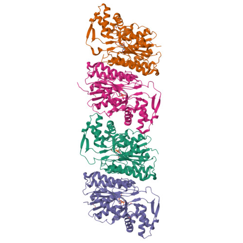
Crystal Structure of DNA Polymerase III beta subunit from Elizabethkingia anophelis
DeBouver, N.D., Abendroth, J., Lorimer, D.D., Horanyi, P.S., Edwards, T.E.To be published.
Experimental Data Snapshot
Starting Model: in silico
View more details
Entity ID: 1 | |||||
|---|---|---|---|---|---|
| Molecule | Chains | Sequence Length | Organism | Details | Image |
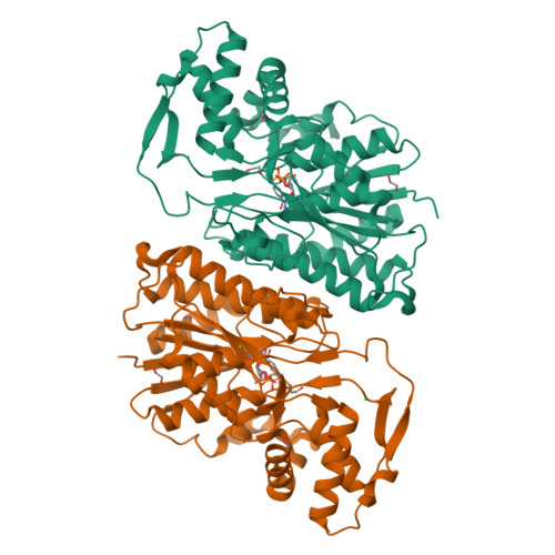
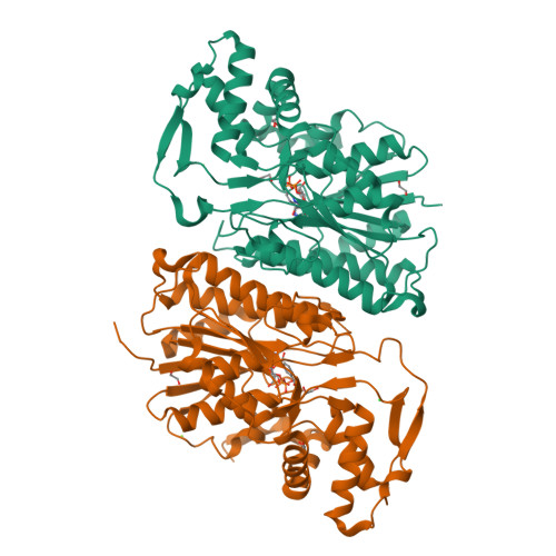
| dTDP-glucose 4,6-dehydratase | 367 | Elizabethkingia anophelis NUHP1 | Mutation(s): 0 Gene Names: BD94_3274 EC: 4.2.1.46 | 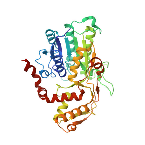 | |
UniProt | |||||
Find proteins for A0A077ELH2 (Elizabethkingia anophelis NUHP1) Explore A0A077ELH2 Go to UniProtKB: A0A077ELH2 | |||||
Entity Groups | |||||
| Sequence Clusters | 30% Identity50% Identity70% Identity90% Identity95% Identity100% Identity | ||||
| UniProt Group | A0A077ELH2 | ||||
Sequence AnnotationsExpand | |||||
| |||||
| Ligands 4 Unique | |||||
|---|---|---|---|---|---|
| ID | Chains | Name / Formula / InChI Key | 2D Diagram | 3D Interactions | |
| NAD (Subject of Investigation/LOI) Query on NAD | F [auth A], K [auth B], Q [auth C], W [auth D] | NICOTINAMIDE-ADENINE-DINUCLEOTIDE C21 H27 N7 O14 P2 BAWFJGJZGIEFAR-NNYOXOHSSA-N |  | ||
| EDO Query on EDO | E [auth A] G [auth A] H [auth A] J [auth B] L [auth B] | 1,2-ETHANEDIOL C2 H6 O2 LYCAIKOWRPUZTN-UHFFFAOYSA-N |  | ||
| CL Query on CL | I [auth A], N [auth B], T [auth C], Z [auth D] | CHLORIDE ION Cl VEXZGXHMUGYJMC-UHFFFAOYSA-M |  | ||
| MG Query on MG | O [auth C], U [auth D] | MAGNESIUM ION Mg JLVVSXFLKOJNIY-UHFFFAOYSA-N |  | ||
| Length ( Å ) | Angle ( ˚ ) |
|---|---|
| a = 66.74 | α = 90 |
| b = 84.62 | β = 101.18 |
| c = 130.87 | γ = 90 |
| Software Name | Purpose |
|---|---|
| XDS | data reduction |
| XSCALE | data scaling |
| PHENIX | refinement |
| PDB_EXTRACT | data extraction |
| PHASER | phasing |
| Funding Organization | Location | Grant Number |
|---|---|---|
| Not funded | -- |
RCSB PDB is hosted by

RCSB PDB is a member of the