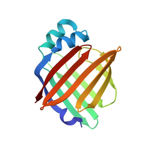
Lipid exchange in crystal-confined fatty acid binding proteins: X-ray evidence and molecular dynamics explanation.
Alvarez, H.A., Cousido-Siah, A., Espinosa, Y.R., Podjarny, A., Carlevaro, C.M., Howard, E.(2023) Proteins 91: 1525-1534
- PubMed: 37462340
- DOI: https://doi.org/10.1002/prot.26546
- Primary Citation of Related Structures:
8GEW - PubMed Abstract:
Fatty acid binding proteins (FABPs) are responsible for the long-chain fatty acids (FAs) transport inside the cell. However, despite the years, since their structure is known and the many studies published, there is no definitive answer about the stages of the lipid entry-exit mechanism. Their structure forms a -barrel of 10 anti-parallel strands with a cap in a helix-turn-helix motif, and there is some consensus on the role of the so-called portal region, involving the second -helix from the cap ( 2), C- D, and E- F turns in FAs exchange. To test the idea of a lid that opens, we performed a soaking experiment on an h-FABP crystal in which the cap is part of the packing contacts, and its movement is strongly restricted. Even in these conditions, we observed the replacement of palmitic acid by 2-Bromohexadecanoic acid (Br-palmitic acid). Our MD simulations reveal a two-step lipid entry process: (i) The travel of the lipid head through the cavity in the order of tens of nanoseconds, and (ii) The accommodation of its hydrophobic tail in hundreds to thousands of nanoseconds. We observed this even in the cases in which the FAs enter the cavity by their tail. During this process, the FAs do not follow a single trajectory, but multiple ones through which they get into the protein cavity. Thanks to the complementary views between experiment and simulation, we can give an approach to a mechanistic view of the exchange process.
Organizational Affiliation:
Instituto de Fisica de Liquidos y Sistemas Biologicos (UNLP-CONICET), Buenos Aires, Argentina.


