8IC6

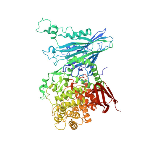
exo-beta-D-arabinanase ExoMA2 from Microbacterium arabinogalactanolyticum in complex with Tris


Experimental Data Snapshot

  • Method: X-RAY DIFFRACTION
  • Resolution: 1.75 Å
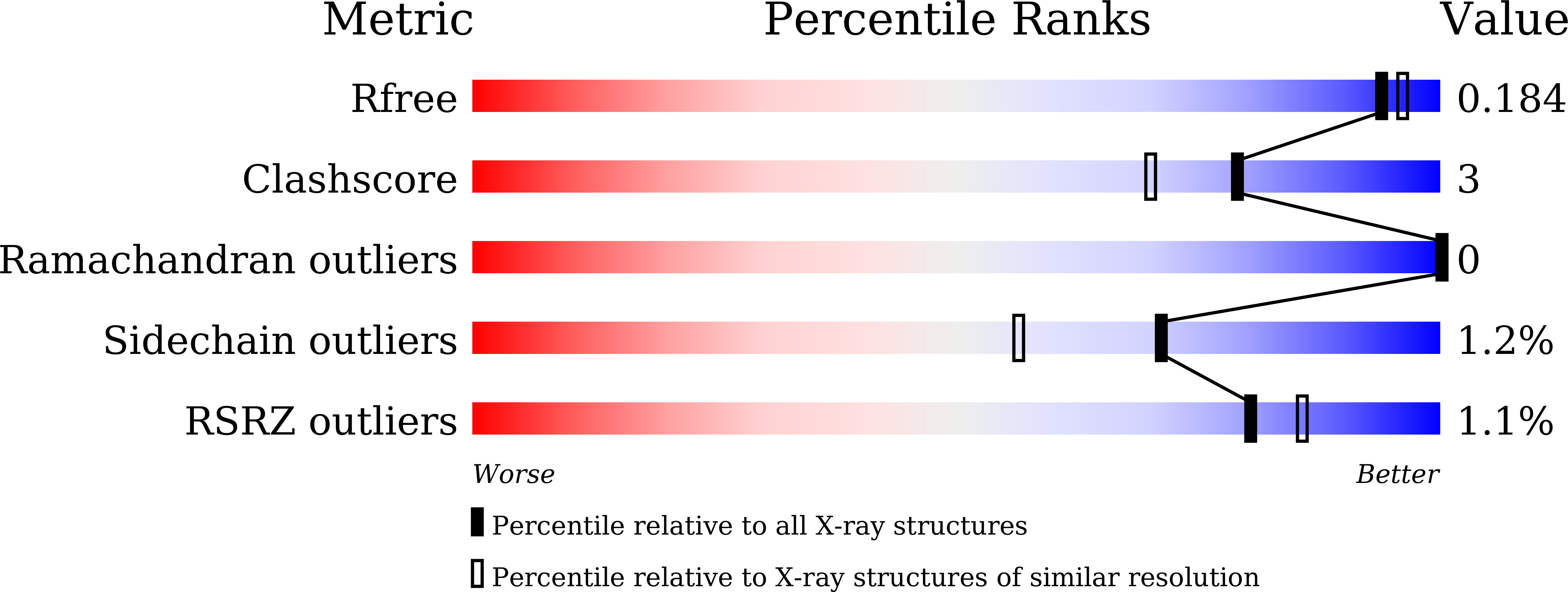
  • R-Value Free: 0.174 
  • R-Value Work: 0.138 
  • R-Value Observed: 0.139 

Starting Model: in silico
View more details

wwPDB Validation   3D Report Full Report


This is version 1.1 of the entry. See complete history


Literature

Identification and characterization of endo-alpha-, exo-alpha-, and exo-beta-D-arabinofuranosidases degrading lipoarabinomannan and arabinogalactan of mycobacteria.

Shimokawa, M.Ishiwata, A.Kashima, T.Nakashima, C.Li, J.Fukushima, R.Sawai, N.Nakamori, M.Tanaka, Y.Kudo, A.Morikami, S.Iwanaga, N.Akai, G.Shimizu, N.Arakawa, T.Yamada, C.Kitahara, K.Tanaka, K.Ito, Y.Fushinobu, S.Fujita, K.

(2023) Nat Commun 14: 5803-5803

  • DOI: https://doi.org/10.1038/s41467-023-41431-2
  • Primary Citation of Related Structures:  
    8HHV, 8IC1, 8IC6, 8IC7, 8IC8

  • PubMed Abstract: 

    The cell walls of pathogenic and acidophilic bacteria, such as Mycobacterium tuberculosis and Mycobacterium leprae, contain lipoarabinomannan and arabinogalactan. These components are composed of D-arabinose, the enantiomer of the typical L-arabinose found in plants. The unique glycan structures of mycobacteria contribute to their ability to evade mammalian immune responses. In this study, we identified four enzymes (two GH183 endo-D-arabinanases, GH172 exo-α-D-arabinofuranosidase, and GH116 exo-β-D-arabinofuranosidase) from Microbacterium arabinogalactanolyticum. These enzymes completely degraded the complex D-arabinan core structure of lipoarabinomannan and arabinogalactan in a concerted manner. Furthermore, through biochemical characterization using synthetic substrates and X-ray crystallography, we elucidated the mechanisms of substrate recognition and anomer-retaining hydrolysis for the α- and β-D-arabinofuranosidic bonds in both endo- and exo-mode reactions. The discovery of these D-arabinan-degrading enzymes, along with the understanding of their structural basis for substrate specificity, provides valuable resources for investigating the intricate glycan architecture of mycobacterial cell wall polysaccharides and their contribution to pathogenicity.


  • Organizational Affiliation

    Faculty of Agriculture, Kagoshima University, Kagoshima, 890-0065, Japan.


Macromolecules
Find similar proteins by:  (by identity cutoff)  |  3D Structure
Entity ID: 1
MoleculeChains Sequence LengthOrganismDetailsImage
exo-beta-D-arabinanaseA [auth B],
B [auth A]
867Microbacterium arabinogalactanolyticumMutation(s): 0 
Gene Names: MIAR_33170
Entity Groups  
Sequence Clusters30% Identity50% Identity70% Identity90% Identity95% Identity100% Identity
Sequence Annotations
Expand
  • Reference Sequence
Small Molecules
Ligands 5 Unique
IDChains Name / Formula / InChI Key2D Diagram3D Interactions
TRS
Query on TRS

Download Ideal Coordinates CCD File 
K [auth B],
W [auth A]
2-AMINO-2-HYDROXYMETHYL-PROPANE-1,3-DIOL
C4 H12 N O3
LENZDBCJOHFCAS-UHFFFAOYSA-O
MPD
Query on MPD

Download Ideal Coordinates CCD File 
L [auth B],
N [auth B],
X [auth A],
Y [auth A]
(4S)-2-METHYL-2,4-PENTANEDIOL
C6 H14 O2
SVTBMSDMJJWYQN-YFKPBYRVSA-N
MLI
Query on MLI

Download Ideal Coordinates CCD File 
M [auth B]MALONATE ION
C3 H2 O4
OFOBLEOULBTSOW-UHFFFAOYSA-L
CL
Query on CL

Download Ideal Coordinates CCD File 
V [auth A]CHLORIDE ION
Cl
VEXZGXHMUGYJMC-UHFFFAOYSA-M
MG
Query on MG

Download Ideal Coordinates CCD File 
C [auth B]
D [auth B]
E [auth B]
F [auth B]
G [auth B]
C [auth B],
D [auth B],
E [auth B],
F [auth B],
G [auth B],
H [auth B],
I [auth B],
J [auth B],
O [auth A],
P [auth A],
Q [auth A],
R [auth A],
S [auth A],
T [auth A],
U [auth A]
MAGNESIUM ION
Mg
JLVVSXFLKOJNIY-UHFFFAOYSA-N
Experimental Data & Validation

Experimental Data

  • Method: X-RAY DIFFRACTION
  • Resolution: 1.75 Å
  • R-Value Free: 0.174 
  • R-Value Work: 0.138 
  • R-Value Observed: 0.139 
  • Space Group: P 1 21 1
Unit Cell:
Length ( Å )Angle ( ˚ )
a = 66.552α = 90
b = 97.511β = 100.55
c = 139.978γ = 90
Software Package:
Software NamePurpose
REFMACrefinement
XDSdata reduction
Aimlessdata scaling
MOLREPphasing

Structure Validation

View Full Validation Report


Entry History & Funding Information

Deposition Data


Funding OrganizationLocationGrant Number
Japan Society for the Promotion of Science (JSPS)Japan15H02443
Japan Society for the Promotion of Science (JSPS)Japan26660083
Japan Society for the Promotion of Science (JSPS)Japan24380053

Revision History  (Full details and data files)

  • Version 1.0: 2023-08-16
    Type: Initial release
  • Version 1.1: 2023-09-27
    Changes: Database references