8PMU

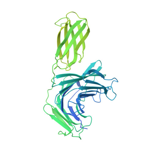
Crystal structure of the HC7 apo form


Experimental Data Snapshot

  • Method: X-RAY DIFFRACTION
  • Resolution: 1.75 Å
  • R-Value Free: 0.231 
  • R-Value Work: 0.191 
  • R-Value Observed: 0.193 

Starting Model: in silico
View more details

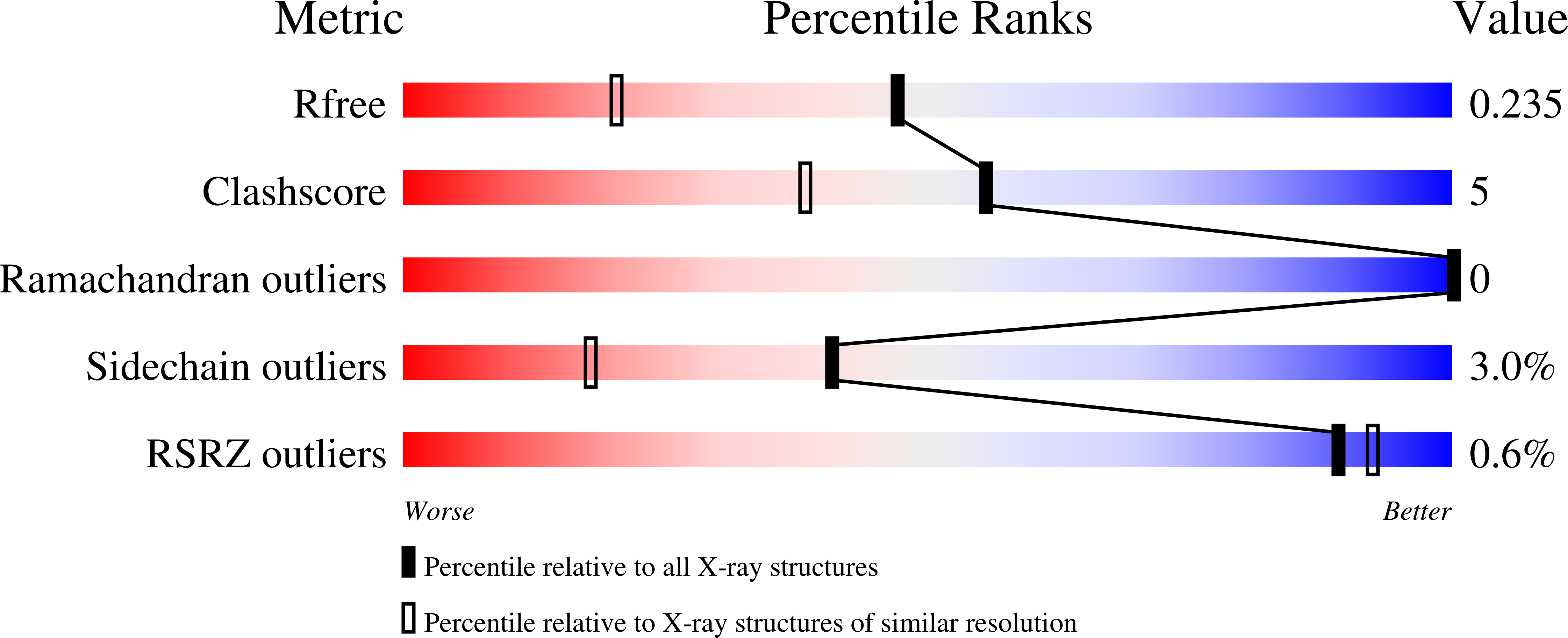
wwPDB Validation   3D Report Full Report


This is version 1.0 of the entry. See complete history


Literature

A family of di-glutamate mucin-degrading enzymes that bridges glycan hydrolases and peptidases

Narimatsu, Y.Bull, C.Taleb, V.Liao, Q.Companon, I.Sanchez-Navarro, D.Durbesson, F.Vincentelli, R.Hansen, L.Corzana, F.Rovira, C.Henrissat, B.Clausen, H.Joshi, H.J.Hurtado-Guerrero, R.

(2024) Nat Catal 


Macromolecules
Find similar proteins by:  (by identity cutoff)  |  3D Structure
Entity ID: 1
MoleculeChains Sequence LengthOrganismDetailsImage
DUF3472 domain-containing protein659Bacillus cereusMutation(s): 0 
Gene Names: GE376_31780
Entity Groups  
Sequence Clusters30% Identity50% Identity70% Identity90% Identity95% Identity100% Identity
Sequence Annotations
Expand
  • Reference Sequence
Small Molecules
Ligands 1 Unique
IDChains Name / Formula / InChI Key2D Diagram3D Interactions
GOL
Query on GOL

Download Ideal Coordinates CCD File 
B [auth A]GLYCEROL
C3 H8 O3
PEDCQBHIVMGVHV-UHFFFAOYSA-N
Experimental Data & Validation

Experimental Data

  • Method: X-RAY DIFFRACTION
  • Resolution: 1.75 Å
  • R-Value Free: 0.231 
  • R-Value Work: 0.191 
  • R-Value Observed: 0.193 
  • Space Group: P 41 21 2
Unit Cell:
Length ( Å )Angle ( ˚ )
a = 93.854α = 90
b = 93.854β = 90
c = 87.287γ = 90
Software Package:
Software NamePurpose
REFMACrefinement
XDSdata reduction
SCALAdata scaling
PHASERphasing

Structure Validation

View Full Validation Report


Entry History & Funding Information

Deposition Data


Funding OrganizationLocationGrant Number
Spanish Ministry of Science, Innovation, and UniversitiesSpain--

Revision History  (Full details and data files)

  • Version 1.0: 2024-03-06
    Type: Initial release