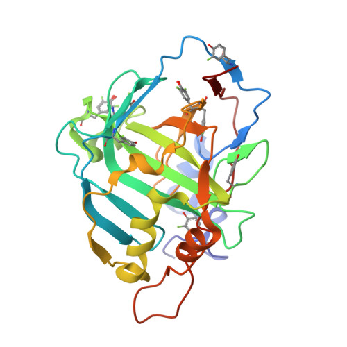
8Q0C

Human carbonic anhydrase II containing 3-fluorotyrosine


Experimental Data Snapshot

  • Method: X-RAY DIFFRACTION
  • Resolution: 1.30 Å
  • R-Value Free: 0.197 
  • R-Value Work: 0.165 
  • R-Value Observed: 0.167 

Starting Model: experimental
View more details

wwPDB Validation   3D Report Full Report


Ligand Structure Quality Assessment 


This is version 1.1 of the entry. See complete history


Literature

Controlling the incorporation of fluorinated amino acids in human cells and its structural impact.

Costantino, A.Pham, L.B.T.Barbieri, L.Calderone, V.Ben-Nissan, G.Sharon, M.Banci, L.Luchinat, E.

(2024) Protein Sci 33: e4910-e4910

  • DOI: https://doi.org/10.1002/pro.4910
  • Primary Citation of Related Structures:  
    8P6U, 8PHL, 8Q0C

  • PubMed Abstract: 

    Fluorinated aromatic amino acids (FAAs) are promising tools when studying protein structure and dynamics by NMR spectroscopy. The incorporation FAAs in mammalian expression systems has been introduced only recently. Here, we investigate the effects of FAAs incorporation in proteins expressed in human cells, focusing on the probability of incorporation and its consequences on the 19 F NMR spectra. By combining 19 F NMR, direct MS and x-ray crystallography, we demonstrate that the probability of FAA incorporation is only a function of the FAA concentration in the expression medium and is a pure stochastic phenomenon. In contrast with the MS data, the x-ray structures of carbonic anhydrase II reveal that while the 3D structure is not affected, certain positions lack fluorine, suggesting that crystallization selectively excludes protein molecules featuring subtle conformational modifications. This study offers a predictive model of the FAA incorporation efficiency and provides a framework for controlling protein fluorination in mammalian expression systems.


  • Organizational Affiliation

    CERM - Magnetic Resonance Center, Università degli Studi di Firenze, Sesto Fiorentino, Italy.


Macromolecules
Find similar proteins by:  (by identity cutoff)  |  3D Structure
Entity ID: 1
MoleculeChains Sequence LengthOrganismDetailsImage
Carbonic anhydrase 2258Homo sapiensMutation(s): 0 
Gene Names: CA2
EC: 4.2.1.1 (PDB Primary Data), 4.2.1.69 (PDB Primary Data)
UniProt & NIH Common Fund Data Resources
Find proteins for P00918 (Homo sapiens)
Explore P00918 
Go to UniProtKB:  P00918
PHAROS:  P00918
GTEx:  ENSG00000104267 
Entity Groups  
Sequence Clusters30% Identity50% Identity70% Identity90% Identity95% Identity100% Identity
UniProt GroupP00918
Sequence Annotations
Expand
  • Reference Sequence
Small Molecules
Modified Residues  1 Unique
IDChains TypeFormula2D DiagramParent
YOF
Query on YOF
A
L-PEPTIDE LINKINGC9 H10 F N O3TYR
Experimental Data & Validation

Experimental Data

  • Method: X-RAY DIFFRACTION
  • Resolution: 1.30 Å
  • R-Value Free: 0.197 
  • R-Value Work: 0.165 
  • R-Value Observed: 0.167 
  • Space Group: P 1 21 1
Unit Cell:
Length ( Å )Angle ( ˚ )
a = 41.99α = 90
b = 41.14β = 104.2
c = 71.21γ = 90
Software Package:
Software NamePurpose
PHENIXrefinement
XDSdata reduction
XSCALEdata scaling
MOLREPphasing

Structure Validation

View Full Validation Report


Ligand Structure Quality Assessment 


Entry History & Funding Information

Deposition Data


Funding OrganizationLocationGrant Number
Italian Ministry of EducationItaly--

Revision History  (Full details and data files)

  • Version 1.0: 2024-03-06
    Type: Initial release
  • Version 1.1: 2024-11-13
    Changes: Structure summary