1DGK | pdb_00001dgk

MUTANT MONOMER OF RECOMBINANT HUMAN HEXOKINASE TYPE I WITH GLUCOSE AND ADP IN THE ACTIVE SITE


Experimental Data Snapshot

  • Method: X-RAY DIFFRACTION
  • Resolution: 2.80 Å
  • R-Value Free: 
    0.308 (Depositor) 
  • R-Value Work: 
    0.258 (Depositor), 0.220 (DCC) 
  • R-Value Observed: 
    0.260 (Depositor) 

wwPDB Validation   3D Report Full Report


Ligand Structure Quality Assessment 

Created with Raphaël 2.3.0Worse 01 BetterLigand structure goodness of fit to experimental dataBest fitted ADPClick on this verticalbar to view detailsBest fitted GLCClick on this verticalbar to view details

This is version 1.6 of the entry. See complete history


Literature

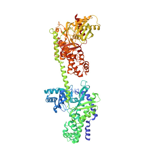
Crystal structures of mutant monomeric hexokinase I reveal multiple ADP binding sites and conformational changes relevant to allosteric regulation.

Aleshin, A.E.Kirby, C.Liu, X.Bourenkov, G.P.Bartunik, H.D.Fromm, H.J.Honzatko, R.B.

(2000) J Mol Biology 296: 1001-1015

  • DOI: https://doi.org/10.1006/jmbi.1999.3494
  • Primary Citation of Related Structures:  
    1CZA, 1DGK

  • PubMed Abstract: 

    Hexokinase I, the pacemaker of glycolysis in brain tissue, is composed of two structurally similar halves connected by an alpha-helix. The enzyme dimerizes at elevated protein concentrations in solution and in crystal structures; however, almost all published data reflect the properties of a hexokinase I monomer in solution. Crystal structures of mutant forms of recombinant human hexokinase I, presented here, reveal the enzyme monomer for the first time. The mutant hexokinases bind both glucose 6-phosphate and glucose with high affinity to their N and C-terminal halves, and ADP, also with high affinity, to a site near the N terminus of the polypeptide chain. Exposure of the monomer crystals to ADP in the complete absence of glucose 6-phosphate reveals a second binding site for adenine nucleotides at the putative active site (C-half), with conformational changes extending 15 A to the contact interface between the N and C-halves. The structures reveal distinct conformational states for the C-half and a rigid-body rotation of the N-half, as possible elements of a structure-based mechanism for allosteric regulation of catalysis.


  • Organizational Affiliation

    Department of Biochemistry Biophysics and Molecular Biology, Iowa State University, Ames, IA 50011, USA.


Macromolecules
Find similar proteins by:  (by identity cutoff)  |  3D Structure
Entity ID: 1
MoleculeChains Sequence LengthOrganismDetailsImage
HEXOKINASE TYPE IA [auth N]917Homo sapiensMutation(s): 4 
EC: 2.7.1.1
UniProt & NIH Common Fund Data Resources
Find proteins for P19367 (Homo sapiens)
Explore P19367 
Go to UniProtKB:  P19367
PHAROS:  P19367
GTEx:  ENSG00000156515 
Entity Groups  
Sequence Clusters30% Identity50% Identity70% Identity90% Identity95% Identity100% Identity
UniProt GroupP19367
Sequence Annotations
Expand
  • Reference Sequence
Experimental Data & Validation

Experimental Data

  • Method: X-RAY DIFFRACTION
  • Resolution: 2.80 Å
  • R-Value Free:  0.308 (Depositor) 
  • R-Value Work:  0.258 (Depositor), 0.220 (DCC) 
  • R-Value Observed: 0.260 (Depositor) 
Space Group: P 21 21 2
Unit Cell:
Length ( Å )Angle ( ˚ )
a = 145.78α = 90
b = 146.83β = 90
c = 58.59γ = 90
Software Package:
Software NamePurpose
AMoREphasing
REFMACrefinement
DENZOdata reduction
SCALEPACKdata scaling

Structure Validation

View Full Validation Report


Ligand Structure Quality Assessment 

Created with Raphaël 2.3.0Worse 01 BetterLigand structure goodness of fit to experimental dataBest fitted ADPClick on this verticalbar to view detailsBest fitted GLCClick on this verticalbar to view details

Entry History 

Deposition Data

Revision History  (Full details and data files)

  • Version 1.0: 2000-03-08
    Type: Initial release
  • Version 1.1: 2008-04-27
    Changes: Version format compliance
  • Version 1.2: 2011-07-13
    Changes: Version format compliance
  • Version 1.3: 2018-01-31
    Changes: Database references
  • Version 1.4: 2020-07-29
    Type: Remediation
    Reason: Carbohydrate remediation
    Changes: Data collection, Database references, Derived calculations, Structure summary
  • Version 1.5: 2021-11-03
    Changes: Database references, Structure summary
  • Version 1.6: 2024-02-07
    Changes: Data collection