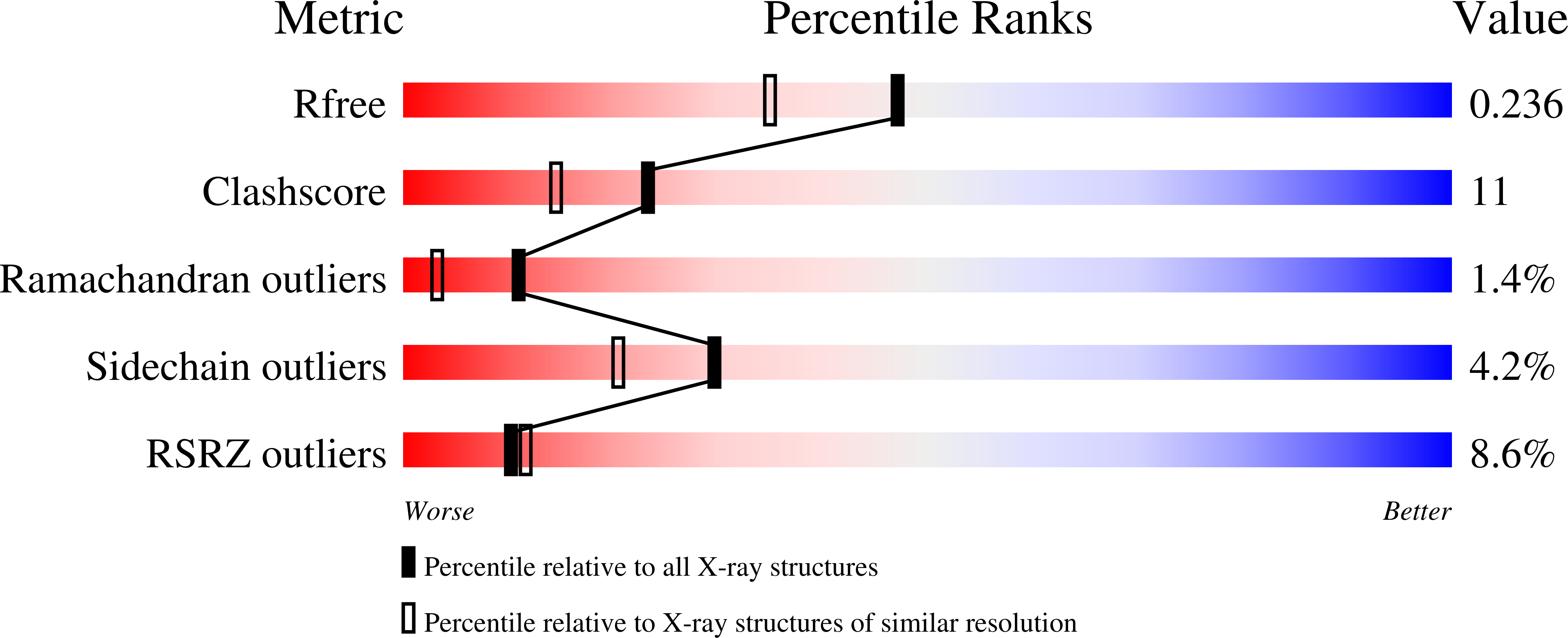
Residue R216 and catalytic efficiency of a murine class alpha glutathione S-transferase toward benzo[a]pyrene 7(R),8(S)-diol 9(S), 10(R)-epoxide.
Gu, Y., Singh, S.V., Ji, X.(2000) Biochemistry 39: 12552-12557
- PubMed: 11027134
- DOI: https://doi.org/10.1021/bi001396u
- Primary Citation of Related Structures:
1F3A, 1F3B - PubMed Abstract:
Murine class alpha glutathione S-transferase A1-1 (mGSTA1-1), unlike mammalian class alpha GSTs, is the most efficient in the glutathione (GSH) conjugation of the ultimate carcinogenic metabolite of benzo[a]pyrene, (+)-anti-7,8-dihydroxy-9,10-oxy-7,8,9, 10-tetrahydrobenzo[a]pyrene [(+)-anti-BPDE] [Hu, X., Srivastava, S. K., Xia, H., Awasthi, Y. C., and Singh, S. V. (1996) J. Biol. Chem. 271, 32684-32688]. Here, we report the crystal structures of mGSTA1-1 in complex with GSH and with the GSH conjugate of (+)-anti-BPDE (GSBpd) at 1.9 and 2.0 A resolution, respectively. Both crystals belong to monoclinic space group C2 with one dimer in the asymmetric unit. The structures reveal that, within one subunit, the GSH moiety interacts with residues Y8, R14, K44, Q53, V54, Q66, and T67, whereas the hydrophobic moiety of GSBpd interacts with the side chains of F9, R14, M207, A215, R216, F219, and I221. In addition, the GSH moiety interacts with D100 and R130 from the other subunit across the dimer interface. The structural comparison between mGSTA1-1.GSH and mGSTA1-1.GSBpd reveals significant conformational differences. The movement of helix alpha9 brings the residues on the helix into direct interaction with the product. Most noticeable are the positional displacement and conformational change of R216, one of the residues located in helix alpha9. The side chain of R216, which points away from the H-site in the mGSTA1-1.GSH complex, probes into the active site and becomes parallel with the aromatic ring system of GSBpd. Moreover, the guanidinium group of R216 shifts approximately 8 A and forms a strong hydrogen bond with the C8 hydroxyl group of GSBpd, suggesting that the electrostatic assistance provided by the guanidinium group facilitates the ring-opening reaction of (+)-anti-BPDE. The structure of mGSTA1-1. GSBpd is also compared with those of hGSTP1-1[V104,A113].GSBpd, hGSPA1-1.S-benzylglutathione, and mGSTA4-4. 4-S-glutathionyl-5-pentyltetrahydrofuran-2-ol. The comparison provides further evidence that supports the functional roles of R216 and helix alpha9. The lack of mobility of helix alpha9 and/or the lack of electrostatic assistance from R216 may be responsible for the relatively lower activity of hGSTA1-1, mGSTA4-4, and hGSTP1-1 toward (+)-anti-BPDE.
Organizational Affiliation:
Program in Structural Biology, National Cancer Institute-Frederick Cancer Research and Development Center, Frederick, Maryland 21702, USA.


