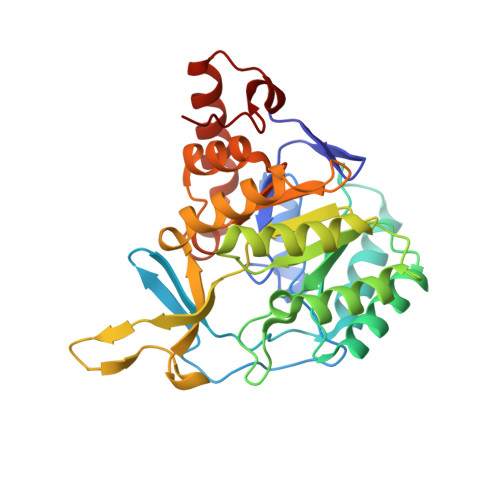
1JRB

The P56A mutant of Lactococcus lactis dihydroorotate dehydrogenase A


Experimental Data Snapshot

  • Method: X-RAY DIFFRACTION
  • Resolution: 1.90 Å
  • R-Value Free: 0.236 
  • R-Value Work: 0.196 

Starting Model: experimental
View more details

wwPDB Validation   3D Report Full Report


Ligand Structure Quality Assessment 


This is version 1.5 of the entry. See complete history


Literature

Lactococcus lactis dihydroorotate dehydrogenase A mutants reveal important facets of the enzymatic function

Norager, S.Arent, S.Bjornberg, O.Ottosen, M.Lo Leggio, L.Jensen, K.F.Larsen, S.

(2003) J Biol Chem 278: 28812-28822

  • DOI: https://doi.org/10.1074/jbc.M303767200
  • Primary Citation of Related Structures:  
    1JQV, 1JQX, 1JRB, 1JRC, 1JUB, 1JUE, 1OVD

  • PubMed Abstract: 

    Dihydroorotate dehydrogenases (DHODs) are flavoenzymes catalyzing the oxidation of (S)-dihydroorotate to orotate in the biosynthesis of UMP, the precursor of all other pyrimidine nucleotides. On the basis of sequence, DHODs can be divided into two classes, class 1, further divided in subclasses 1A and 1B, and class 2. This division corresponds to differences in cellular location and the nature of the electron acceptor. Herein we report a study of Lactococcus lactis DHODA, a representative of the class 1A enzymes. Based on the DHODA structure we selected seven residues that are highly conserved between both main classes of DHODs as well as three residues representing surface charges close to the active site for site-directed mutagenesis. The availability of both kinetic and structural data on the mutant enzymes allowed us to define the roles individual structural segments play in catalysis. We have also structurally proven the presence of an open active site loop in DHODA and obtained information about the interactions that control movements of loops around the active site. Furthermore, in one mutant structure we observed differences between the two monomers of the dimer, confirming an apparent asymmetry between the two substrate binding sites that was indicated by the kinetic results.


  • Organizational Affiliation

    Centre for Crystallographic Studies, University of Copenhagen, Universitetsparken 5, DK-2100 Copenhagen, Denmark.


Macromolecules
Find similar proteins by:  (by identity cutoff)  |  3D Structure
Entity ID: 1
MoleculeChains Sequence LengthOrganismDetailsImage
dihydroorotate dehydrogenase A
A, B
311Lactococcus lactisMutation(s): 1 
EC: 1.3.3.1 (PDB Primary Data), 1.3.98.1 (UniProt)
UniProt
Find proteins for A2RJT9 (Lactococcus lactis subsp. cremoris (strain MG1363))
Explore A2RJT9 
Go to UniProtKB:  A2RJT9
Entity Groups  
Sequence Clusters30% Identity50% Identity70% Identity90% Identity95% Identity100% Identity
UniProt GroupA2RJT9
Sequence Annotations
Expand
  • Reference Sequence
Experimental Data & Validation

Experimental Data

  • Method: X-RAY DIFFRACTION
  • Resolution: 1.90 Å
  • R-Value Free: 0.236 
  • R-Value Work: 0.196 
  • Space Group: P 1 21 1
Unit Cell:
Length ( Å )Angle ( ˚ )
a = 53.074α = 90
b = 108.822β = 103.92
c = 66.453γ = 90
Software Package:
Software NamePurpose
DENZOdata reduction
SCALEPACKdata scaling
AMoREphasing
REFMACrefinement

Structure Validation

View Full Validation Report


Ligand Structure Quality Assessment 


Entry History 

Deposition Data

Revision History  (Full details and data files)

  • Version 1.0: 2003-09-09
    Type: Initial release
  • Version 1.1: 2008-04-27
    Changes: Version format compliance
  • Version 1.2: 2011-07-13
    Changes: Version format compliance
  • Version 1.3: 2018-05-23
    Changes: Data collection
  • Version 1.4: 2021-11-10
    Changes: Database references, Derived calculations
  • Version 1.5: 2023-10-25
    Changes: Data collection, Refinement description