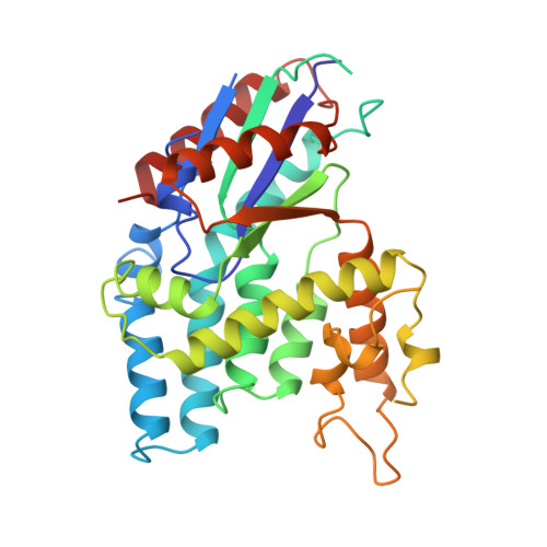
Exploring the active site of herpes simplex virus type-1 thymidine kinase by X-ray crystallography of complexes with aciclovir and other ligands.
Champness, J.N., Bennett, M.S., Wien, F., Visse, R., Summers, W.C., Herdewijn, P., de Clerq, E., Ostrowski, T., Jarvest, R.L., Sanderson, M.R.(1998) Proteins 32: 350-361
- PubMed: 9715911
- DOI: https://doi.org/10.1002/(sici)1097-0134(19980815)32:3<350::aid-prot10>3.0.co;2-8
- Primary Citation of Related Structures:
1KI2, 1KI3, 1KI4, 1KI6, 1KI7, 1KI8, 1KIM - PubMed Abstract:
Antiherpes therapies are principally targeted at viral thymidine kinases and utilize nucleoside analogs, the triphosphates of which are inhibitors of viral DNA polymerase or result in toxic effects when incorporated into DNA. The most frequently used drug, aciclovir (Zovirax), is a relatively poor substrate for thymidine kinase and high-resolution structural information on drugs and other molecules binding to the target is therefore important for the design of novel and more potent chemotherapy, both in antiherpes treatment and in gene therapy systems where thymidine kinase is expressed. Here, we report for the first time the binary complexes of HSV-1 thymidine kinase (TK) with the drug molecules aciclovir and penciclovir, determined by X-ray crystallography at 2.37 A resolution. Moreover, from new data at 2.14 A resolution, the refined structure of the complex of TK with its substrate deoxythymidine (R = 0.209 for 96% of all data) now reveals much detail concerning substrate and solvent interactions with the enzyme. Structures of the complexes of TK with four halogen-containing substrate analogs have also been solved, to resolutions better than 2.4 A. The various TK inhibitors broadly fall into three groups which together probe the space of the enzyme active site in a manner that no one molecule does alone, so giving a composite picture of active site interactions that can be exploited in the design of novel compounds.
Organizational Affiliation:
Division of Biomedical Sciences, Randall Institute, King's College, London, United Kingdom.


