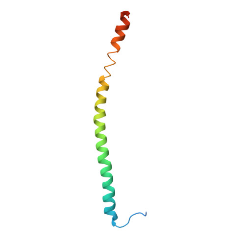
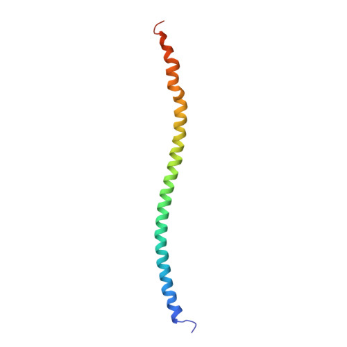
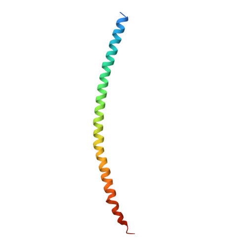
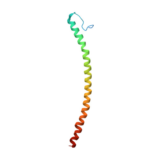
X-ray structure of a neuronal complexin-SNARE complex from squid.
Bracher, A., Kadlec, J., Betz, H., Weissenhorn, W.(2002) J Biological Chem 277: 26517-26523
- PubMed: 12004067
- DOI: https://doi.org/10.1074/jbc.M203460200
- Primary Citation of Related Structures:
1L4A - PubMed Abstract:
Nerve terminals release neurotransmitters from vesicles into the synaptic cleft upon transient increases in intracellular Ca(2+). This exocytotic process requires the formation of trans SNARE complexes and is regulated by accessory proteins including the complexins. Here we report the crystal structure of a squid core complexin-SNARE complex at 2.95-A resolution. A helical segment of complexin binds in anti-parallel fashion to the four-helix bundle of the core SNARE complex and interacts at its C terminus with syntaxin and synaptobrevin around the ionic zero layer of the SNARE complex. We propose that this structure is part of a multiprotein fusion machinery that regulates vesicle fusion at a late pre-fusion stage. Accordingly, Ca(2+) may initiate membrane fusion by acting directly or indirectly on complexin, thus allowing the conformational transitions of the trans SNARE complex that are thought to drive membrane fusion.
- European Molecular Biology Laboratory, 6 rue Jules Horowitz, 38042 Grenoble, France.
Organizational Affiliation: