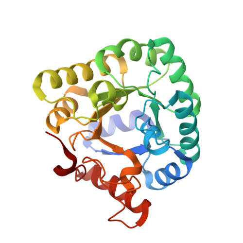
1M3U

Crystal Structure of Ketopantoate Hydroxymethyltransferase complexed the Product Ketopantoate


Experimental Data Snapshot

  • Method: X-RAY DIFFRACTION
  • Resolution: 1.80 Å
  • R-Value Free: 0.193 
  • R-Value Work: 0.152 
  • R-Value Observed: 0.153 

Starting Model: experimental
View more details

wwPDB Validation   3D Report Full Report


This is version 1.5 of the entry. See complete history


Literature

Structure of E. coli Ketopantoate Hydroxymethyl Transferase Complexed with Ketopantoate and Mg(2+), Solved by Locating 160 Selenomethionine Sites.

von Delft, F.Inoue, T.Saldanha, S.A.Ottenhof, H.H.Schmitzberger, F.Birch, L.M.Dhanaraj, V.Witty, M.Smith, A.G.Blundell, T.L.Abell, C.

(2003) Structure 11: 985-996

  • DOI: https://doi.org/10.1016/s0969-2126(03)00158-8
  • Primary Citation of Related Structures:  
    1M3U

  • PubMed Abstract: 

    We report the crystal structure of E. coli ketopantoate hydroxymethyltransferase (KPHMT) at 1.9 A resolution, in complex with its product, ketopantoate. KPHMT catalyzes the first step in the biosynthesis of pantothenate (vitamin B(5)), the precursor of coenzyme A and the acyl carrier protein cofactor. The structure of the decameric enzyme was solved by multiwavelength anomalous dispersion to locate 160 selenomethionine sites and phase 560 kDa of protein, making it the largest structure solved by this approach. KPHMT adopts the (betaalpha)(8) barrel fold and is a member of the phosphoenolpyruvate/pyruvate superfamily. The active site contains a ketopantoate bidentately coordinated to Mg(2+). Similar binding is likely for the substrate, alpha-ketoisovalerate, orienting the C3 for deprotonation.


  • Organizational Affiliation

    Department of Biochemistry, 80 Tennis Court Road, Cambridge CB2 1GA, United Kingdom.


Macromolecules
Find similar proteins by:  (by identity cutoff)  |  3D Structure
Entity ID: 1
MoleculeChains Sequence LengthOrganismDetailsImage
3-methyl-2-oxobutanoate hydroxymethyltransferase
A, B, C, D, E
A, B, C, D, E, F, G, H, I, J
264Escherichia coliMutation(s): 0 
Gene Names: PANB
EC: 2.1.2.11
UniProt
Find proteins for P31057 (Escherichia coli (strain K12))
Explore P31057 
Go to UniProtKB:  P31057
Entity Groups  
Sequence Clusters30% Identity50% Identity70% Identity90% Identity95% Identity100% Identity
UniProt GroupP31057
Sequence Annotations
Expand
  • Reference Sequence
Small Molecules
Ligands 2 Unique
IDChains Name / Formula / InChI Key2D Diagram3D Interactions
KPL
Query on KPL

Download Ideal Coordinates CCD File 
BA [auth I]
DA [auth J]
L [auth A]
N [auth B]
P [auth C]
BA [auth I],
DA [auth J],
L [auth A],
N [auth B],
P [auth C],
R [auth D],
T [auth E],
V [auth F],
X [auth G],
Z [auth H]
KETOPANTOATE
C6 H10 O4
PKVVTUWHANFMQC-UHFFFAOYSA-N
MG
Query on MG

Download Ideal Coordinates CCD File 
AA [auth I]
CA [auth J]
K [auth A]
M [auth B]
O [auth C]
AA [auth I],
CA [auth J],
K [auth A],
M [auth B],
O [auth C],
Q [auth D],
S [auth E],
U [auth F],
W [auth G],
Y [auth H]
MAGNESIUM ION
Mg
JLVVSXFLKOJNIY-UHFFFAOYSA-N
Experimental Data & Validation

Experimental Data

  • Method: X-RAY DIFFRACTION
  • Resolution: 1.80 Å
  • R-Value Free: 0.193 
  • R-Value Work: 0.152 
  • R-Value Observed: 0.153 
  • Space Group: P 1 21 1
Unit Cell:
Length ( Å )Angle ( ˚ )
a = 86.074α = 90
b = 157.17β = 97.44
c = 100.181γ = 90
Software Package:
Software NamePurpose
MOSFLMdata reduction
SCALAdata scaling
SnBphasing
SHARPphasing
REFMACrefinement
CCP4data scaling

Structure Validation

View Full Validation Report


Entry History 

Deposition Data

Revision History  (Full details and data files)

  • Version 1.0: 2003-07-22
    Type: Initial release
  • Version 1.1: 2008-04-28
    Changes: Version format compliance
  • Version 1.2: 2011-07-13
    Changes: Advisory, Version format compliance
  • Version 1.3: 2017-10-11
    Changes: Refinement description
  • Version 1.4: 2024-02-14
    Changes: Data collection, Database references, Derived calculations, Refinement description
  • Version 1.5: 2024-04-03
    Changes: Refinement description