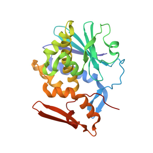
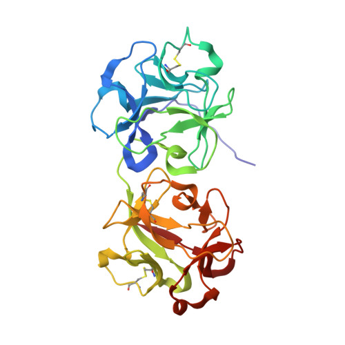
1OQL | pdb_00001oql

Mistletoe Lectin I from Viscum album complexed with galactose


Experimental Data Snapshot

  • Method: X-RAY DIFFRACTION
  • Resolution: 3.00 Å
  • R-Value Free: 
    0.240 (Depositor), 0.230 (DCC) 
  • R-Value Work: 
    0.199 (Depositor), 0.190 (DCC) 
  • R-Value Observed: 
    0.201 (Depositor) 

Starting Model: experimental
View more details

wwPDB Validation   3D Report Full Report


Ligand Structure Quality Assessment 

Created with Raphaël 2.3.0Worse 01 BetterLigand structure goodness of fit to experimental dataBest fitted NAGClick on this verticalbar to view detailsBest fitted GALClick on this verticalbar to view details

This is version 2.2 of the entry. See complete history


Literature

Crystal structure at 3 A of mistletoe lectin I, a dimeric type-II ribosome-inactivating protein, complexed with galactose

Niwa, H.Tonevitsky, A.G.Agapov, I.I.Saward, S.Pfuller, U.Palmer, R.A.

(2003) Eur J Biochem 270: 2739-2749

  • DOI: https://doi.org/10.1046/j.1432-1033.2003.03646.x
  • Primary Citation of Related Structures:  
    1OQL

  • PubMed Abstract: 

    The X-ray structure of mistletoe lectin I (MLI), a type-II ribosome-inactivating protein (RIP), cocrystallized with galactose is described. The model was refined at 3.0 A resolution to an R-factor of 19.9% using 21 899 reflections, with Rfree 24.0%. MLI forms a homodimer (A-B)2 in the crystal, as it does in solution at high concentration. The dimer is formed through contacts between the N-terminal domains of two B-chains involving weak polar and non-polar interactions. Consequently, the overall arrangement of sugar-binding sites in MLI differs from those in monomeric type-II RIPs: two N-terminal sugar-binding sites are 15 A apart on one side of the dimer, and two C-terminal sugar-binding sites are 87 A apart on the other side. Galactose binding is achieved by common hydrogen bonds for the two binding sites via hydroxy groups 3-OH and 4-OH and hydrophobic contact by an aromatic ring. In addition, at the N-terminal site 2-OH forms hydrogen bonds with Asp27 and Lys41, and at the C-terminal site 3-OH and 6-OH undergo water-mediated interactions and C5 has a hydrophobic contact. MLI is a galactose-specific lectin and shows little affinity for N-acetylgalactosamine. The reason for this is discussed. Structural differences among the RIPs investigated in this study (their quaternary structures, location of sugar-binding sites, and fine sugar specificities of their B-chains, which could have diverged through evolution from a two-domain protein) may affect the binding sites, and consequently the cellular transport processes and biological responses of these toxins.


  • Organizational Affiliation

    School of Crystallography, Birkbeck College, University of London, Malet Street, London WC1E 7HX, UK. h.niwa@mail.cryst.bbk.ac.uk


Macromolecules
Find similar proteins by:  (by identity cutoff)  |  3D Structure
Entity ID: 1
MoleculeChains Sequence LengthOrganismDetailsImage
MISTLETOE LECTIN I249Viscum albumMutation(s): 0 
EC: 3.2.2.22
UniProt
Find proteins for P81446 (Viscum album)
Explore P81446 
Go to UniProtKB:  P81446
Entity Groups  
Sequence Clusters30% Identity50% Identity70% Identity90% Identity95% Identity100% Identity
UniProt GroupP81446
Glycosylation
Glycosylation Sites: 1
Sequence Annotations
Expand
  • Reference Sequence
Find similar proteins by:  (by identity cutoff)  |  3D Structure
Entity ID: 2
MoleculeChains Sequence LengthOrganismDetailsImage
MISTLETOE LECTIN I263Viscum albumMutation(s): 0 
UniProt
Find proteins for P81446 (Viscum album)
Explore P81446 
Go to UniProtKB:  P81446
Entity Groups  
Sequence Clusters30% Identity50% Identity70% Identity90% Identity95% Identity100% Identity
UniProt GroupP81446
Glycosylation
Glycosylation Sites: 3
Sequence Annotations
Expand
  • Reference Sequence
Oligosaccharides

Help

Entity ID: 3
MoleculeChains Length2D Diagram Glycosylation3D Interactions
2-acetamido-2-deoxy-beta-D-glucopyranose-(1-4)-2-acetamido-2-deoxy-beta-D-glucopyranose
C, D
2N-Glycosylation
Glycosylation Resources
GlyTouCan:  G42666HT
GlyCosmos:  G42666HT
GlyGen:  G42666HT
Experimental Data & Validation

Experimental Data

  • Method: X-RAY DIFFRACTION
  • Resolution: 3.00 Å
  • R-Value Free:  0.240 (Depositor), 0.230 (DCC) 
  • R-Value Work:  0.199 (Depositor), 0.190 (DCC) 
  • R-Value Observed: 0.201 (Depositor) 
Space Group: P 65 2 2
Unit Cell:
Length ( Å )Angle ( ˚ )
a = 107.65α = 90
b = 107.65β = 90
c = 311.92γ = 120
Software Package:
Software NamePurpose
CNSrefinement
DENZOdata reduction
CCP4data scaling
AMoREphasing

Structure Validation

View Full Validation Report


Ligand Structure Quality Assessment 

Created with Raphaël 2.3.0Worse 01 BetterLigand structure goodness of fit to experimental dataBest fitted NAGClick on this verticalbar to view detailsBest fitted GALClick on this verticalbar to view details

Entry History 

Deposition Data

Revision History  (Full details and data files)

  • Version 1.0: 2003-07-01
    Type: Initial release
  • Version 1.1: 2008-04-29
    Changes: Version format compliance
  • Version 1.2: 2011-07-13
    Changes: Derived calculations, Version format compliance
  • Version 2.0: 2020-07-29
    Type: Remediation
    Reason: Carbohydrate remediation
    Changes: Atomic model, Data collection, Derived calculations, Structure summary
  • Version 2.1: 2023-10-25
    Changes: Data collection, Database references, Refinement description, Structure summary
  • Version 2.2: 2024-10-23
    Changes: Structure summary