1OUT

TROUT HEMOGLOBIN I


Experimental Data Snapshot

  • Method: X-RAY DIFFRACTION
  • Resolution: 2.30 Å
  • R-Value Free: 0.247 
  • R-Value Work: 0.162 

wwPDB Validation   3D Report Full Report


This is version 1.2 of the entry. See complete history


Literature

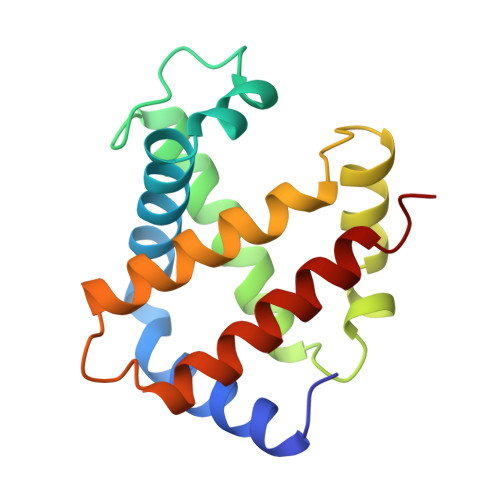
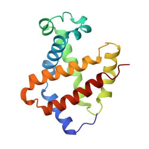
The crystal structures of trout Hb I in the deoxy and carbonmonoxy forms.

Tame, J.R.Wilson, J.C.Weber, R.E.

(1996) J Mol Biol 259: 749-760

  • DOI: https://doi.org/10.1006/jmbi.1996.0355
  • Primary Citation of Related Structures:  
    1OUT, 1OUU

  • PubMed Abstract: 

    We have determined the X-ray crystallographic structure of trout Hb I in both the deoxy and carbonmonoxy forms to resolution limits of 2.3 angstroms and 2.5 angstroms, respectively. The overall fold of the molecule is highly similar to that of human HbA despite the low level of sequence identity between these proteins. Trout Hb I is unusual in displaying almost no pH dependence of oxygen binding affinity, and (at most) very weak interactions with heterotropic effector ligands such as organic phosphates. Comparison of the two quaternary states of the protein indicates how such effects are minimised and how the low-affinity T state of the protein is stabilised in the absence of heterotropic interactions.


  • Organizational Affiliation

    Department of Chemistry, University of York, Helsington, UK.


Macromolecules
Find similar proteins by:  (by identity cutoff)  |  3D Structure
Entity ID: 1
MoleculeChains Sequence LengthOrganismDetailsImage
HEMOGLOBIN I143Oncorhynchus mykissMutation(s): 0 
UniProt
Find proteins for P02019 (Oncorhynchus mykiss)
Explore P02019 
Go to UniProtKB:  P02019
Entity Groups  
Sequence Clusters30% Identity50% Identity70% Identity90% Identity95% Identity100% Identity
UniProt GroupP02019
Sequence Annotations
Expand
  • Reference Sequence
Find similar proteins by:  (by identity cutoff)  |  3D Structure
Entity ID: 2
MoleculeChains Sequence LengthOrganismDetailsImage
HEMOGLOBIN I146Oncorhynchus mykissMutation(s): 0 
UniProt
Find proteins for P02142 (Oncorhynchus mykiss)
Explore P02142 
Go to UniProtKB:  P02142
Entity Groups  
Sequence Clusters30% Identity50% Identity70% Identity90% Identity95% Identity100% Identity
UniProt GroupP02142
Sequence Annotations
Expand
  • Reference Sequence
Small Molecules
Ligands 1 Unique
IDChains Name / Formula / InChI Key2D Diagram3D Interactions
HEM
Query on HEM

Download Ideal Coordinates CCD File 
C [auth A],
D [auth B]
PROTOPORPHYRIN IX CONTAINING FE
C34 H32 Fe N4 O4
KABFMIBPWCXCRK-RGGAHWMASA-L
Experimental Data & Validation

Experimental Data

  • Method: X-RAY DIFFRACTION
  • Resolution: 2.30 Å
  • R-Value Free: 0.247 
  • R-Value Work: 0.162 
  • Space Group: P 65 2 2
Unit Cell:
Length ( Å )Angle ( ˚ )
a = 63.28α = 90
b = 63.28β = 90
c = 312.74γ = 120
Software Package:
Software NamePurpose
PROLSQrefinement
MOSFLMdata reduction

Structure Validation

View Full Validation Report


Entry History 

Deposition Data

Revision History  (Full details and data files)

  • Version 1.0: 1997-01-11
    Type: Initial release
  • Version 1.1: 2008-03-24
    Changes: Version format compliance
  • Version 1.2: 2011-07-13
    Changes: Version format compliance