1QMZ

PHOSPHORYLATED CDK2-CYCLYIN A-SUBSTRATE PEPTIDE COMPLEX


Experimental Data Snapshot

  • Method: X-RAY DIFFRACTION
  • Resolution: 2.20 Å
  • R-Value Free: 0.280 
  • R-Value Work: 0.220 

Starting Model: other
View more details

wwPDB Validation   3D Report Full Report


This is version 1.4 of the entry. See complete history


Literature

The Structural Basis for Specificity of Substrate and Recruitment Peptides for Cyclin-Dependent Kinases

Brown, N.R.Noble, M.E.Endicott, J.A.Johnson, L.N.

(1999) Nat Cell Biol 1: 438

  • DOI: https://doi.org/10.1038/15674
  • Primary Citation of Related Structures:  
    1QMZ

  • PubMed Abstract: 

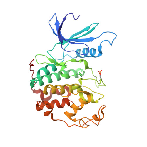
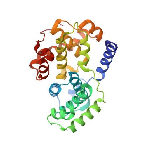
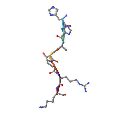
    Progression through the eukaryotic cell cycle is driven by the orderly activation of cyclin-dependent kinases (CDKs). For activity, CDKs require association with a cyclin and phosphorylation by a separate protein kinase at a conserved threonine residue (T160 in CDK2). Here we present the structure of a complex consisting of phosphorylated CDK2 and cyclin A together with an optimal peptide substrate, HHASPRK. This structure provides an explanation for the specificity of CDK2 towards the proline that follows the phosphorylatable serine of the substrate peptide, and the requirement for the basic residue in the P+3 position of the substrate. We also present the structure of phosphorylated CDK2 plus cyclin A3 in complex with residues 658-668 from the CDK2 substrate p107. These residues include the RXL motif required to target p107 to cyclins. This structure explains the specificity of the RXL motif for cyclins.


  • Organizational Affiliation

    Laboratory of Molecular Biophysics and Oxford Centre for Molecular Sciences, Department of Biochemistry, University of Oxford, Rex Richards Building, South Parks Road, Oxford OX1 3QU, UK.


Macromolecules
Find similar proteins by:  (by identity cutoff)  |  3D Structure
Entity ID: 1
MoleculeChains Sequence LengthOrganismDetailsImage
CELL DIVISION PROTEIN KINASE 2
A, C
299Homo sapiensMutation(s): 0 
EC: 2.7.1 (PDB Primary Data), 2.7.11.22 (UniProt)
UniProt & NIH Common Fund Data Resources
Find proteins for P24941 (Homo sapiens)
Explore P24941 
Go to UniProtKB:  P24941
PHAROS:  P24941
GTEx:  ENSG00000123374 
Entity Groups  
Sequence Clusters30% Identity50% Identity70% Identity90% Identity95% Identity100% Identity
UniProt GroupP24941
Sequence Annotations
Expand
  • Reference Sequence
Find similar proteins by:  (by identity cutoff)  |  3D Structure
Entity ID: 2
MoleculeChains Sequence LengthOrganismDetailsImage
G2/MITOTIC-SPECIFIC CYCLIN A
B, D
259Homo sapiensMutation(s): 0 
UniProt & NIH Common Fund Data Resources
Find proteins for P20248 (Homo sapiens)
Explore P20248 
Go to UniProtKB:  P20248
PHAROS:  P20248
GTEx:  ENSG00000145386 
Entity Groups  
Sequence Clusters30% Identity50% Identity70% Identity90% Identity95% Identity100% Identity
UniProt GroupP20248
Sequence Annotations
Expand
  • Reference Sequence

Find similar proteins by:  Sequence   |   3D Structure  

Entity ID: 3
MoleculeChains Sequence LengthOrganismDetailsImage
SUBSTRATE PEPTIDE
E, F
7Homo sapiensMutation(s): 0 
Sequence Annotations
Expand
  • Reference Sequence
Small Molecules
Experimental Data & Validation

Experimental Data

  • Method: X-RAY DIFFRACTION
  • Resolution: 2.20 Å
  • R-Value Free: 0.280 
  • R-Value Work: 0.220 
  • Space Group: P 21 21 2
Unit Cell:
Length ( Å )Angle ( ˚ )
a = 152.6α = 90
b = 163.7β = 90
c = 73.3γ = 90
Software Package:
Software NamePurpose
REFMACrefinement
MOSFLMdata reduction
SCALAdata scaling
AMoREphasing

Structure Validation

View Full Validation Report


Entry History 

Deposition Data

Revision History  (Full details and data files)

  • Version 1.0: 1999-12-14
    Type: Initial release
  • Version 1.1: 2011-05-08
    Changes: Version format compliance
  • Version 1.2: 2011-07-13
    Changes: Version format compliance
  • Version 1.3: 2024-05-01
    Changes: Data collection, Database references, Derived calculations, Refinement description
  • Version 1.4: 2024-10-16
    Changes: Structure summary