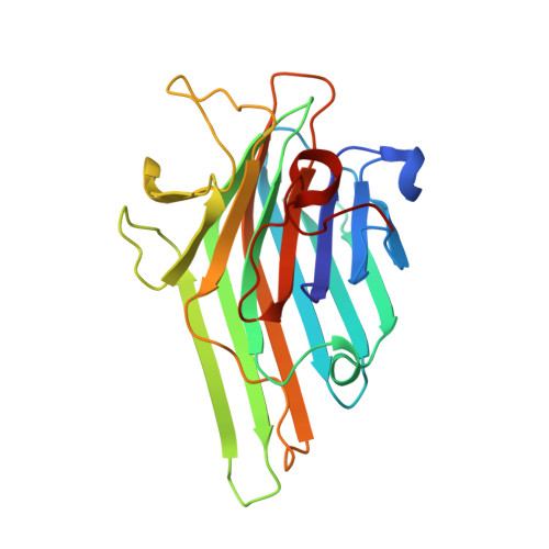
1QNY

X-ray refinement of D2O soaked crystal of concanavalin A


Experimental Data Snapshot

  • Method: X-RAY DIFFRACTION
  • Resolution: 1.80 Å
  • R-Value Free: 0.198 
  • R-Value Work: 0.168 
  • R-Value Observed: 0.168 

wwPDB Validation   3D Report Full Report


This is version 1.3 of the entry. See complete history


Literature

Direct Determination of the Positions of Deuterium Atoms of Bound Water in Concanavalin a by Neutron Laue Crystallography

Habash, J.Raftery, J.Nuttall, R.Price, H.J.Lehmann, M.S.Wilkinson, C.Kalb(Gilboa), A.J.Helliwell, J.R.

(2000) Acta Crystallogr D Biol Crystallogr 56: 541

  • DOI: https://doi.org/10.1107/s0907444900002353
  • Primary Citation of Related Structures:  
    1C57, 1QNY

  • PubMed Abstract: 

    The correct positions of the deuterium (D) atoms of many of the bound waters in the protein concanavalin A are revealed by neutron Laue diffraction. The approach includes cases where these water D atoms show enough mobility to render them invisible even to ultra-high resolution synchrotron-radiation X-ray crystallography. The positions of the bound water H atoms calculated on the basis of chemical and energetic considerations are often incorrect. The D-atom positions for the water molecules in the Mn-, Ca- and sugar-binding sites of concanavalin A are described in detail.


  • Organizational Affiliation

    Section of Structural Chemistry, Department of Chemistry, University of Manchester, Manchester M13 9PL, England.


Macromolecules
Find similar proteins by:  (by identity cutoff)  |  3D Structure
Entity ID: 1
MoleculeChains Sequence LengthOrganismDetailsImage
CONCANAVALIN A237Canavalia ensiformisMutation(s): 0 
UniProt
Find proteins for P02866 (Canavalia ensiformis)
Explore P02866 
Go to UniProtKB:  P02866
Entity Groups  
Sequence Clusters30% Identity50% Identity70% Identity90% Identity95% Identity100% Identity
UniProt GroupP02866
Sequence Annotations
Expand
  • Reference Sequence
Small Molecules
Ligands 2 Unique
IDChains Name / Formula / InChI Key2D Diagram3D Interactions
MN
Query on MN

Download Ideal Coordinates CCD File 
B [auth A]MANGANESE (II) ION
Mn
WAEMQWOKJMHJLA-UHFFFAOYSA-N
CA
Query on CA

Download Ideal Coordinates CCD File 
C [auth A]CALCIUM ION
Ca
BHPQYMZQTOCNFJ-UHFFFAOYSA-N
Experimental Data & Validation

Experimental Data

  • Method: X-RAY DIFFRACTION
  • Resolution: 1.80 Å
  • R-Value Free: 0.198 
  • R-Value Work: 0.168 
  • R-Value Observed: 0.168 
  • Space Group: I 2 2 2
Unit Cell:
Length ( Å )Angle ( ˚ )
a = 89.11α = 90
b = 87.58β = 90
c = 63.26γ = 90
Software Package:
Software NamePurpose
X-PLORrefinement
DENZOdata reduction
SCALEPACKdata scaling
X-PLORphasing

Structure Validation

View Full Validation Report


Entry History 

Deposition Data

Revision History  (Full details and data files)

  • Version 1.0: 2000-04-30
    Type: Initial release
  • Version 1.1: 2011-05-08
    Changes: Version format compliance
  • Version 1.2: 2011-07-13
    Changes: Version format compliance
  • Version 1.3: 2024-05-08
    Changes: Data collection, Database references, Derived calculations, Other