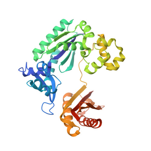
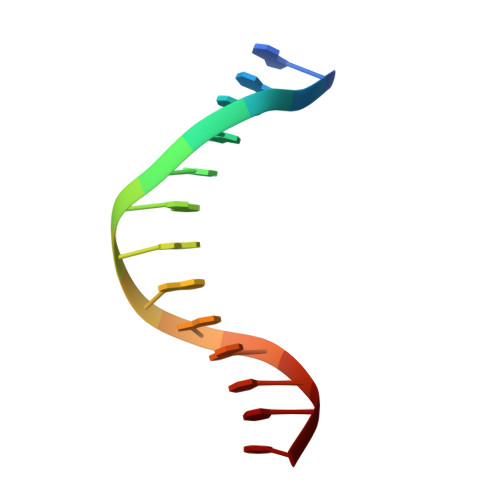
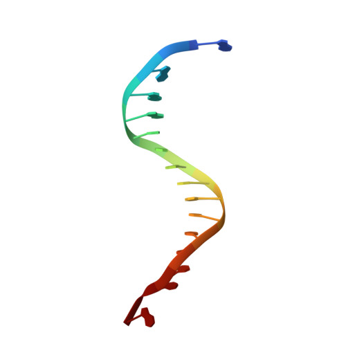
1S0M

Crystal structure of a Benzo[a]pyrene Diol Epoxide adduct in a ternary complex with a DNA polymerase


Experimental Data Snapshot

  • Method: X-RAY DIFFRACTION
  • Resolution: 2.70 Å
  • R-Value Free: 0.246 
  • R-Value Work: 0.207 
  • R-Value Observed: 0.207 

Starting Model: experimental
View more details

wwPDB Validation   3D Report Full Report


Ligand Structure Quality Assessment 


This is version 1.5 of the entry. See complete history


Literature

Crystal structure of a benzo[a]pyrene diol epoxide adduct in a ternary complex with a DNA polymerase.

Ling, H.Sayer, J.M.Plosky, B.S.Yagi, H.Boudsocq, F.Woodgate, R.Jerina, D.M.Yang, W.

(2004) Proc Natl Acad Sci U S A 101: 2265-2269

  • DOI: https://doi.org/10.1073/pnas.0308332100
  • Primary Citation of Related Structures:  
    1S0M

  • PubMed Abstract: 

    The first occupation-associated cancers to be recognized were the sooty warts (cancers of the scrotum) suffered by chimney sweeps in 18th century England. In the 19th century, high incidences of skin cancers were noted among fuel industry workers. By the early 20th century, malignant skin tumors were produced in laboratory animals by repeatedly painting them with coal tar. The culprit in coal tar that induces cancer was finally isolated in 1933 and determined to be benzo[a]pyrene (BP), a polycyclic aromatic hydrocarbon. A residue of fuel and tobacco combustion and frequently ingested by humans, BP is metabolized in mammals to benzo[a]pyrene diol epoxide (BPDE), which forms covalent DNA adducts and induces tumor growth. In the 70 yr since its isolation, BP has been the most studied carcinogen. Yet, there has been no crystal structure of a BPDE DNA adduct. We report here the crystal structure of a BPDE-adenine adduct base-paired with thymine at a template-primer junction and complexed with the lesion-bypass DNA polymerase Dpo4 and an incoming nucleotide. Two conformations of the BPDE, one intercalated between base pairs and another solvent-exposed in the major groove, are observed. The latter conformation, which can be stabilized by organic solvents that reduce the dielectric constant, seems more favorable for DNA replication by Dpo4. These structures also suggest a mechanism by which mutations are generated during replication of DNA containing BPDE adducts.


  • Organizational Affiliation

    Laboratory of Molecular Biology, National Institute of Diabetes and Digestive and Kidney Diseases, National Institute of Child Health and Human Development, National Institutes of Health, Bethesda, MD 20892, USA.


Macromolecules

Find similar proteins by:  (by identity cutoff)  |  3D Structure
Entity ID: 3
MoleculeChains Sequence LengthOrganismDetailsImage
DNA polymerase IVE [auth A],
F [auth B]
352Saccharolobus solfataricus P2Mutation(s): 0 
Gene Names: DPO4
EC: 2.7.7.7
UniProt
Find proteins for Q97W02 (Saccharolobus solfataricus (strain ATCC 35092 / DSM 1617 / JCM 11322 / P2))
Explore Q97W02 
Go to UniProtKB:  Q97W02
Entity Groups  
Sequence Clusters30% Identity50% Identity70% Identity90% Identity95% Identity100% Identity
UniProt GroupQ97W02
Sequence Annotations
Expand
  • Reference Sequence

Find similar nucleic acids by:  Sequence   |   3D Structure  

Entity ID: 1
MoleculeChains LengthOrganismImage
5'-D(*GP*GP*GP*GP*GP*AP*AP*GP*GP*AP*TP*TP*T)-3'A [auth C],
C [auth E]
13N/A
Sequence Annotations
Expand
  • Reference Sequence

Find similar nucleic acids by:  Sequence   |   3D Structure  

Entity ID: 2
MoleculeChains LengthOrganismImage
5'-D(P*AP*TP*AP*AP*AP*TP*CP*CP*TP*TP*CP*CP*CP*CP*CP*A)-3'B [auth D],
D [auth F]
16N/A
Sequence Annotations
Expand
  • Reference Sequence
Small Molecules
Ligands 4 Unique
IDChains Name / Formula / InChI Key2D Diagram3D Interactions
DTP
Query on DTP

Download Ideal Coordinates CCD File 
N [auth A],
R [auth B]
2'-DEOXYADENOSINE 5'-TRIPHOSPHATE
C10 H16 N5 O12 P3
SUYVUBYJARFZHO-RRKCRQDMSA-N
BAP
Query on BAP

Download Ideal Coordinates CCD File 
G [auth D],
I [auth F]
1,2,3-TRIHYDROXY-1,2,3,4-TETRAHYDROBENZO[A]PYRENE
C20 H16 O3
GFANZDFKCCJYRF-NSISKUIASA-N
CA
Query on CA

Download Ideal Coordinates CCD File 
H [auth E]
K [auth A]
L [auth A]
M [auth A]
O [auth B]
H [auth E],
K [auth A],
L [auth A],
M [auth A],
O [auth B],
P [auth B],
Q [auth B]
CALCIUM ION
Ca
BHPQYMZQTOCNFJ-UHFFFAOYSA-N
MG
Query on MG

Download Ideal Coordinates CCD File 
J [auth A]MAGNESIUM ION
Mg
JLVVSXFLKOJNIY-UHFFFAOYSA-N
Experimental Data & Validation

Experimental Data

  • Method: X-RAY DIFFRACTION
  • Resolution: 2.70 Å
  • R-Value Free: 0.246 
  • R-Value Work: 0.207 
  • R-Value Observed: 0.207 
  • Space Group: P 21 21 21
Unit Cell:
Length ( Å )Angle ( ˚ )
a = 100.029α = 90
b = 103.545β = 90
c = 106.127γ = 90
Software Package:
Software NamePurpose
HKL-2000data collection
SCALEPACKdata scaling
CNSrefinement
HKL-2000data reduction
CNSphasing

Structure Validation

View Full Validation Report


Ligand Structure Quality Assessment 


Entry History 

Deposition Data

Revision History  (Full details and data files)

  • Version 1.0: 2004-03-30
    Type: Initial release
  • Version 1.1: 2008-04-29
    Changes: Version format compliance
  • Version 1.2: 2011-07-13
    Changes: Source and taxonomy, Version format compliance
  • Version 1.3: 2018-04-04
    Changes: Data collection
  • Version 1.4: 2019-11-20
    Changes: Derived calculations
  • Version 1.5: 2023-08-23
    Changes: Data collection, Database references, Derived calculations, Refinement description