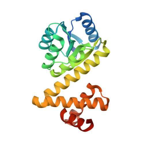
1SD5

Crystal structure of Rv1626


Experimental Data Snapshot

  • Method: X-RAY DIFFRACTION
  • Resolution: 1.68 Å
  • R-Value Free: 0.216 
  • R-Value Work: 0.171 
  • R-Value Observed: 0.173 

wwPDB Validation   3D Report Full Report


This is version 1.3 of the entry. See complete history


Literature

The Crystal and Solution Structure of a Putative Transcriptional Antiterminator from Mycobacterium tuberculosis.

Morth, J.P.Feng, V.Perry, L.J.Svergun, D.I.Tucker, P.A.

(2004) Structure 12: 1595-1605

  • DOI: https://doi.org/10.1016/j.str.2004.06.018
  • Primary Citation of Related Structures:  
    1S8N, 1SD5

  • PubMed Abstract: 

    We describe the crystal structure of Rv1626 from Mycobacterium tuberculosis at 1.48 A resolution and the corresponding solution structure determined from small angle X-ray scattering. The N-terminal domain shows structural homology to the receiver domains found in bacterial two-component systems. The C-terminal domain has high structural homology to a recently discovered RNA binding domain involved in transcriptional antitermination. The molecule in solution was found to be monomeric as it is in the crystal, but in solution it undergoes a conformational change that is triggered by changes in ionic strength. This is the first structure that links the phosphorylation cascade of the two-component systems with the antitermination event in the transcriptional machinery. Rv1626 belongs to a family of proteins, which we propose calling phosphorylation-dependent transcriptional antitermination regulators, so far only found in bacteria, and includes NasT, a protein from the assimilatory nitrate/nitrite reductase operon of Azetobacter vinelandii.


  • Organizational Affiliation

    EMBL Hamburg Outstation, c/o DESY, Notkestrasse 85, Germany.


Macromolecules
Find similar proteins by:  (by identity cutoff)  |  3D Structure
Entity ID: 1
MoleculeChains Sequence LengthOrganismDetailsImage
putative antiterminator205Mycobacterium tuberculosis H37RvMutation(s): 0 
Gene Names: rv1626
UniProt
Find proteins for P9WGM3 (Mycobacterium tuberculosis (strain ATCC 25618 / H37Rv))
Explore P9WGM3 
Go to UniProtKB:  P9WGM3
Entity Groups  
Sequence Clusters30% Identity50% Identity70% Identity90% Identity95% Identity100% Identity
UniProt GroupP9WGM3
Sequence Annotations
Expand
  • Reference Sequence
Small Molecules
Ligands 1 Unique
IDChains Name / Formula / InChI Key2D Diagram3D Interactions
IOD
Query on IOD

Download Ideal Coordinates CCD File 
B [auth A]
C [auth A]
D [auth A]
E [auth A]
F [auth A]
B [auth A],
C [auth A],
D [auth A],
E [auth A],
F [auth A],
G [auth A],
H [auth A],
I [auth A],
J [auth A],
K [auth A],
L [auth A],
M [auth A],
N [auth A],
O [auth A],
P [auth A]
IODIDE ION
I
XMBWDFGMSWQBCA-UHFFFAOYSA-M
Experimental Data & Validation

Experimental Data

  • Method: X-RAY DIFFRACTION
  • Resolution: 1.68 Å
  • R-Value Free: 0.216 
  • R-Value Work: 0.171 
  • R-Value Observed: 0.173 
  • Space Group: P 43 21 2
Unit Cell:
Length ( Å )Angle ( ˚ )
a = 44.558α = 90
b = 44.558β = 90
c = 180.965γ = 90
Software Package:
Software NamePurpose
REFMACrefinement
DENZOdata reduction
SCALEPACKdata scaling
SOLVEphasing

Structure Validation

View Full Validation Report


Entry History 

Deposition Data

Revision History  (Full details and data files)

  • Version 1.0: 2004-09-21
    Type: Initial release
  • Version 1.1: 2008-04-29
    Changes: Version format compliance
  • Version 1.2: 2011-07-13
    Changes: Source and taxonomy, Version format compliance
  • Version 1.3: 2024-02-14
    Changes: Data collection, Database references, Derived calculations