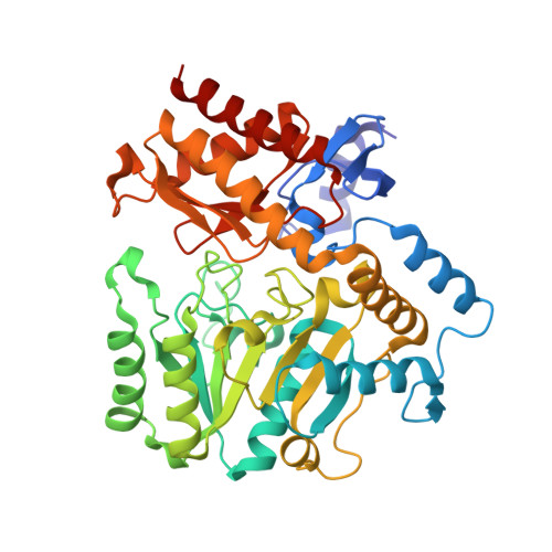
1SZU

The structure of gamma-aminobutyrate aminotransferase mutant: V241A


Experimental Data Snapshot

  • Method: X-RAY DIFFRACTION
  • Resolution: 2.52 Å
  • R-Value Free: 0.202 
  • R-Value Work: 0.154 
  • R-Value Observed: 0.156 

wwPDB Validation   3D Report Full Report


Ligand Structure Quality Assessment 


This is version 1.5 of the entry. See complete history


Literature

Kinetic and Crystallographic Analysis of Active Site Mutants of Escherichia coligamma-Aminobutyrate Aminotransferase.

Liu, W.Peterson, P.E.Langston, J.A.Jin, X.Zhou, X.Fisher, A.J.Toney, M.D.

(2005) Biochemistry 44: 2982-2992

  • DOI: https://doi.org/10.1021/bi048657a
  • Primary Citation of Related Structures:  
    1SZK, 1SZS, 1SZU

  • PubMed Abstract: 

    The E. coli isozyme of gamma-aminobutyrate aminotransferase (GABA-AT) is a tetrameric pyridoxal phosphate-dependent enzyme that catalyzes transamination between primary amines and alpha-keto acids. The roles of the active site residues V241, E211, and I50 in the GABA-AT mechanism have been probed by site-directed mutagenesis. The beta-branched side chain of V241 facilitates formation of external aldimine intermediates with primary amine substrates, while E211 provides charge compensation of R398 selectively in the primary amine half-reaction and I50 forms a hydrophobic lid at the top of the substrate binding site. The structures of the I50Q, V241A, and E211S mutants were solved by X-ray crystallography to resolutions of 2.1, 2.5, and 2.52 A, respectively. The structure of GABA-AT is similar in overall fold and active site structure to that of dialkylglycine decarboxylase, which catalyzes both transamination and decarboxylation half-reactions in its normal catalytic cycle. Therefore, an attempt was made to convert GABA-AT into a decarboxylation-dependent aminotransferase similar to dialkylglycine decarboxylase by systematic mutation of E. coli GABA-AT active site residues. Two of the twelve mutants presented, E211S/I50G/C77K and E211S/I50H/V80D, have approximately 10-fold higher decarboxylation activities than the wild-type enzyme, and the E211S/I50H/V80D has formally changed the reaction specificity to that of a decarboxylase.


  • Organizational Affiliation

    Department of Chemistry, University of California-Davis, Davis, California 95616, USA.


Macromolecules
Find similar proteins by:  (by identity cutoff)  |  3D Structure
Entity ID: 1
MoleculeChains Sequence LengthOrganismDetailsImage
4-aminobutyrate aminotransferase
A, B, C, D
426Escherichia coliMutation(s): 1 
Gene Names: GABTB2662
EC: 2.6.1.19 (PDB Primary Data), 2.6.1.48 (UniProt)
UniProt
Find proteins for P22256 (Escherichia coli (strain K12))
Explore P22256 
Go to UniProtKB:  P22256
Entity Groups  
Sequence Clusters30% Identity50% Identity70% Identity90% Identity95% Identity100% Identity
UniProt GroupP22256
Sequence Annotations
Expand
  • Reference Sequence
Small Molecules
Ligands 4 Unique
IDChains Name / Formula / InChI Key2D Diagram3D Interactions
PMP
Query on PMP

Download Ideal Coordinates CCD File 
CA [auth C],
KA [auth D],
L [auth A],
U [auth B]
4'-DEOXY-4'-AMINOPYRIDOXAL-5'-PHOSPHATE
C8 H13 N2 O5 P
ZMJGSOSNSPKHNH-UHFFFAOYSA-N
PLP
Query on PLP

Download Ideal Coordinates CCD File 
BA [auth C],
JA [auth D],
K [auth A],
T [auth B]
PYRIDOXAL-5'-PHOSPHATE
C8 H10 N O6 P
NGVDGCNFYWLIFO-UHFFFAOYSA-N
SO4
Query on SO4

Download Ideal Coordinates CCD File 
DA [auth D]
E [auth A]
EA [auth D]
F [auth A]
M [auth B]
DA [auth D],
E [auth A],
EA [auth D],
F [auth A],
M [auth B],
N [auth B],
O [auth B],
P [auth B],
V [auth C],
W [auth C],
X [auth C]
SULFATE ION
O4 S
QAOWNCQODCNURD-UHFFFAOYSA-L
EDO
Query on EDO

Download Ideal Coordinates CCD File 
AA [auth C]
FA [auth D]
G [auth A]
GA [auth D]
H [auth A]
AA [auth C],
FA [auth D],
G [auth A],
GA [auth D],
H [auth A],
HA [auth D],
I [auth A],
IA [auth D],
J [auth A],
Q [auth B],
R [auth B],
S [auth B],
Y [auth C],
Z [auth C]
1,2-ETHANEDIOL
C2 H6 O2
LYCAIKOWRPUZTN-UHFFFAOYSA-N
Experimental Data & Validation

Experimental Data

  • Method: X-RAY DIFFRACTION
  • Resolution: 2.52 Å
  • R-Value Free: 0.202 
  • R-Value Work: 0.154 
  • R-Value Observed: 0.156 
  • Space Group: P 32 2 1
Unit Cell:
Length ( Å )Angle ( ˚ )
a = 108.2α = 90
b = 108.2β = 90
c = 301.66γ = 120
Software Package:
Software NamePurpose
XDSdata scaling
SCALEPACKdata scaling
CNSrefinement
XDSdata reduction
CNSphasing

Structure Validation

View Full Validation Report


Ligand Structure Quality Assessment 


Entry History 

Deposition Data

Revision History  (Full details and data files)

  • Version 1.0: 2005-03-01
    Type: Initial release
  • Version 1.1: 2008-04-30
    Changes: Version format compliance
  • Version 1.2: 2011-07-13
    Changes: Non-polymer description, Version format compliance
  • Version 1.3: 2011-11-16
    Changes: Atomic model
  • Version 1.4: 2017-10-11
    Changes: Refinement description
  • Version 1.5: 2021-10-27
    Changes: Database references, Derived calculations