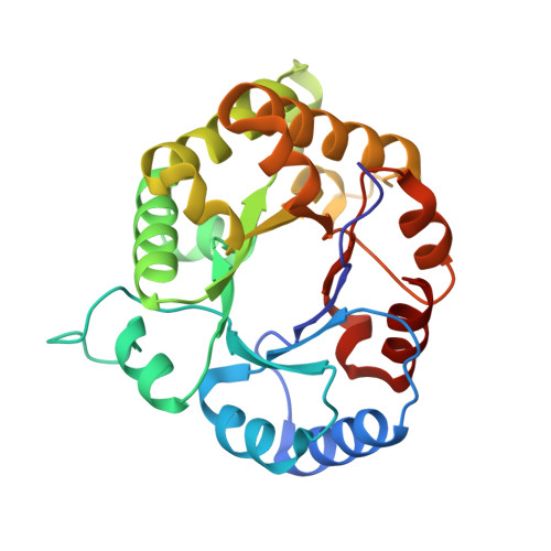
Comparison of the structures and the crystal contacts of trypanosomal triosephosphate isomerase in four different crystal forms.
Kishan, K.V., Zeelen, J.P., Noble, M.E., Borchert, T.V., Wierenga, R.K.(1994) Protein Sci 3: 779-787
- PubMed: 8061607
- DOI: https://doi.org/10.1002/pro.5560030507
- Primary Citation of Related Structures:
1TPE, 1TPF - PubMed Abstract:
Triosephosphate isomerase (TIM) is a dimeric enzyme consisting of 2 identical subunits. Trypanosomal TIM can be crystallized in 4 different spacegroups: P2(1)2(1)2(1), C2(big cell), C2(small cell), and P1. The P1 crystal form only grows in the presence of 1.4 M DMSO; there are 2 DMSO binding sites per subunit. The structures have been refined at a resolution of 1.83 A, 2.10 A, 2.13 A, and 1.80 A, respectively. In the 4 different spacegroups the TIM subunit can be observed in the context of 7 different crystallographic environments. In the C2 cells, the dimer 2-fold axis coincides with a crystallographic 2-fold axis. The similarities and differences of the 7 subunits are discussed. In 6 subunits the flexible loop (loop 6) is open, whereas in the P2(1)2(1)2(1) cell, the flexible loop of subunit 2 is in an almost closed conformation. The crystal contacts in the 4 different crystal forms are predominantly generated by polar residues in loops. A statistical analysis of the residues involved in crystal contacts shows that, in particular, serines are frequently involved in these interactions; 19% of the exposed serines are involved in crystal contacts.
- European Molecular Biology Laboratory, Heidelberg, Germany.
Organizational Affiliation:


