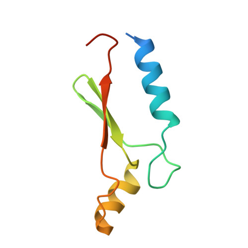
1UW1 | pdb_00001uw1

A Novel ADP- and Zinc-binding fold from function-directed in vitro evolution


Experimental Data Snapshot

  • Method: X-RAY DIFFRACTION
  • Resolution: 1.94 Å
  • R-Value Free: 
    0.228 (Depositor), 0.230 (DCC) 
  • R-Value Work: 
    0.200 (Depositor), 0.210 (DCC) 

wwPDB Validation   3D Report Full Report


Ligand Structure Quality Assessment 

Created with Raphaël 2.3.0Worse 01 BetterLigand structure goodness of fit to experimental dataBest fitted ADPClick on this verticalbar to view details

This is version 1.3 of the entry. See complete history


Literature

A Novel Adp- and Zinc-Binding Fold from Function-Directed in Vitro Evolution

Lo Surdo, P.Walsh, M.A.Sollazzo, M.

(2004) Nat Struct Mol Biol 11: 382

  • DOI: https://doi.org/10.1038/nsmb745
  • Primary Citation of Related Structures:  
    1UW1

  • PubMed Abstract: 

    A great challenge to biologists is to create proteins with novel folds and tailored functions. As an alternative to de novo protein design, we investigated the structure of a randomly generated protein targeted to bind ATP. The crystal structure reveals a novel alpha/beta fold bound to its ligand, representing both the first protein structure derived from in vitro evolution and the first nucleotide-binding protein stabilized by a zinc ion.


  • Organizational Affiliation

    Istituto di Ricerche di Biologia Molecolare (IRBM), Via Pontina Km 30.600, 00040 Pomezia, Roma, Italy. paola_losurdo@merck.com


Macromolecules
Find similar proteins by:  (by identity cutoff)  |  3D Structure
Entity ID: 1
MoleculeChains Sequence LengthOrganismDetailsImage
ARTIFICIAL NUCLEOTIDE BINDING PROTEIN (ANBP)80synthetic constructMutation(s): 0 
Entity Groups  
Sequence Clusters30% Identity50% Identity70% Identity90% Identity95% Identity100% Identity
Sequence Annotations
Expand
  • Reference Sequence
Experimental Data & Validation

Experimental Data

  • Method: X-RAY DIFFRACTION
  • Resolution: 1.94 Å
  • R-Value Free:  0.228 (Depositor), 0.230 (DCC) 
  • R-Value Work:  0.200 (Depositor), 0.210 (DCC) 
Space Group: P 32 2 1
Unit Cell:
Length ( Å )Angle ( ˚ )
a = 71.077α = 90
b = 71.077β = 90
c = 54.883γ = 120
Software Package:
Software NamePurpose
REFMACrefinement
DENZOdata reduction
SCALEPACKdata scaling
SHELXDphasing

Structure Validation

View Full Validation Report


Ligand Structure Quality Assessment 

Created with Raphaël 2.3.0Worse 01 BetterLigand structure goodness of fit to experimental dataBest fitted ADPClick on this verticalbar to view details

Entry History 

Deposition Data

Revision History  (Full details and data files)

  • Version 1.0: 2004-03-26
    Type: Initial release
  • Version 1.1: 2015-10-21
    Changes: Derived calculations, Non-polymer description, Other, Source and taxonomy, Structure summary, Version format compliance
  • Version 1.2: 2019-05-15
    Changes: Data collection, Experimental preparation, Other
  • Version 1.3: 2024-05-08
    Changes: Data collection, Database references, Derived calculations, Other