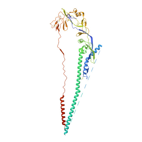
1ZTM

Structure of the Uncleaved Paramyxovirus (hPIV3) Fusion Protein


Experimental Data Snapshot

  • Method: X-RAY DIFFRACTION
  • Resolution: 3.05 Å
  • R-Value Free: 0.264 
  • R-Value Work: 0.217 
  • R-Value Observed: 0.217 

wwPDB Validation   3D Report Full Report


Ligand Structure Quality Assessment 


This is version 1.6 of the entry. See complete history


Literature

Structure of the uncleaved ectodomain of the paramyxovirus (hPIV3) fusion protein

Yin, H.S.Paterson, R.G.Wen, X.Lamb, R.A.Jardetzky, T.S.

(2005) Proc Natl Acad Sci U S A 102: 9288-9293

  • DOI: https://doi.org/10.1073/pnas.0503989102
  • Primary Citation of Related Structures:  
    1ZTM

  • PubMed Abstract: 

    Class I viral fusion proteins share common mechanistic and structural features but little sequence similarity. Structural insights into the protein conformational changes associated with membrane fusion are based largely on studies of the influenza virus hemagglutinin in pre- and postfusion conformations. Here, we present the crystal structure of the secreted, uncleaved ectodomain of the paramyxovirus, human parainfluenza virus 3 fusion (F) protein, a member of the class I viral fusion protein group. The secreted human parainfluenza virus 3 F forms a trimer with distinct head, neck, and stalk regions. Unexpectedly, the structure reveals a six-helix bundle associated with the postfusion form of F, suggesting that the anchor-minus ectodomain adopts a conformation largely similar to the postfusion state. The transmembrane anchor domains of F may therefore profoundly influence the folding energetics that establish and maintain a metastable, prefusion state.


  • Organizational Affiliation

    Department of Biochemistry, Molecular Biology, and Cell Biology, Howard Hughes Medical Institute, Northwestern University, Evanston, IL 60208-3500, USA.


Macromolecules
Find similar proteins by:  (by identity cutoff)  |  3D Structure
Entity ID: 1
MoleculeChains Sequence LengthOrganismDetailsImage
Fusion glycoprotein
A, B, C
490Human respirovirus 3Mutation(s): 1 
Gene Names: F
UniProt
Find proteins for P06828 (Human parainfluenza 3 virus (strain Wash/47885/57))
Explore P06828 
Go to UniProtKB:  P06828
Entity Groups  
Sequence Clusters30% Identity50% Identity70% Identity90% Identity95% Identity100% Identity
UniProt GroupP06828
Glycosylation
Glycosylation Sites: 1
Sequence Annotations
Expand
  • Reference Sequence
Experimental Data & Validation

Experimental Data

  • Method: X-RAY DIFFRACTION
  • Resolution: 3.05 Å
  • R-Value Free: 0.264 
  • R-Value Work: 0.217 
  • R-Value Observed: 0.217 
  • Space Group: P 21 21 21
Unit Cell:
Length ( Å )Angle ( ˚ )
a = 105.56α = 90
b = 122.17β = 90
c = 195.71γ = 90
Software Package:
Software NamePurpose
MAR345data collection
SCALAdata scaling
DMmodel building
CNSrefinement
CCP4data scaling
DMphasing

Structure Validation

View Full Validation Report


Ligand Structure Quality Assessment 


Entry History 

Deposition Data

Revision History  (Full details and data files)

  • Version 1.0: 2005-07-19
    Type: Initial release
  • Version 1.1: 2008-04-30
    Changes: Version format compliance
  • Version 1.2: 2011-07-13
    Changes: Non-polymer description, Version format compliance
  • Version 1.3: 2017-10-11
    Changes: Refinement description
  • Version 1.4: 2020-07-29
    Type: Remediation
    Reason: Carbohydrate remediation
    Changes: Data collection, Database references, Derived calculations, Structure summary
  • Version 1.5: 2021-10-20
    Changes: Database references, Derived calculations, Structure summary
  • Version 1.6: 2024-10-09
    Changes: Data collection, Structure summary