263D | pdb_0000263d

ISOHELICITY AND PHASING IN DRUG-DNA SEQUENCE RECOGNITION: CRYSTAL STRUCTURE OF A TRIS(BENZIMIDAZOLE)-OLIGONUCLEOTIDE COMPLEX


Experimental Data Snapshot

  • Method: X-RAY DIFFRACTION
  • Resolution: 2.20 Å
  • R-Value Work: 
    0.174 (Depositor), 0.180 (DCC) 
  • R-Value Observed: 
    0.174 (Depositor) 

Starting Model: experimental
View more details

wwPDB Validation   3D Report Full Report


Ligand Structure Quality Assessment 

Created with Raphaël 2.3.0Worse 01 BetterLigand structure goodness of fit to experimental dataBest fitted TBZClick on this verticalbar to view details

This is version 1.4 of the entry. See complete history


Literature

Isohelicity and phasing in drug--DNA sequence recognition: crystal structure of a tris(benzimidazole)--oligonucleotide complex.

Clark, G.R.Gray, E.J.Neidle, S.Li, Y.H.Leupin, W.

(1996) Biochemistry 35: 13745-13752

  • DOI: https://doi.org/10.1021/bi960421m
  • Primary Citation of Related Structures:  
    263D

  • PubMed Abstract: 

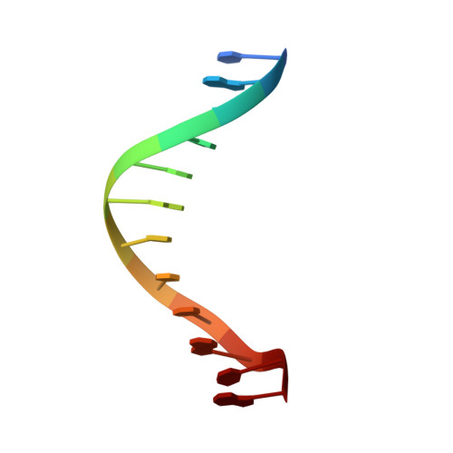
    The crystal structure is reported of a tris(benzimidazole) analogue of the minor-groove drug Hoechst 33258 bound to the sequence d(CGCAAATTTGCG)2. The structure has been refined to an R factor of 17.4% at a resolution of 2.2 A. The ligand covers approximately 7 1/2 base pairs, including the 5'-AAATTT central sequence. This has an exceptionally narrow minor-groove width, together with high propeller twists for individual base pairs. The ligand has a highly twisted structure, with an overall twist of 50 degrees between aromatic rings. All three benzimidazole subunits are in register with the DNA, and there is a symmetric group of six hydrogen bonds between ligand and A.T base-pair edges. By contrast, the ligand does not show an optimal isohelical fit to the DNA. The correct phasing of drug and DNA base pairs is ensured by a number of changes to the DNA such that the central 5'-AAATTT region is slightly unwound relative to the structures of other noncovalent minor-groove drug complexes.


  • Organizational Affiliation

    CRC Biomolecular Structure Unit, Institute of Cancer Research, Sutton, Surrey, U.K.


Macromolecules

Find similar nucleic acids by:  Sequence   |   3D Structure  

Entity ID: 1
MoleculeChains LengthOrganismImage
DNA (5'-D(*CP*GP*CP*AP*AP*AP*TP*TP*TP*GP*CP*G)-3')
A, B
12N/A
Sequence Annotations
Expand
  • Reference Sequence
Small Molecules
Ligands 1 Unique
IDChains Name / Formula / InChI Key2D Diagram3D Interactions
TBZ
Query on TBZ

Download Ideal Coordinates CCD File 
C [auth B]2''-(4-METHOXYPHENYL)-5-(3-AMINO-1-PYRROLIDINYL)-2,5',2',5''-TRI-BENZIMIDAZOLE
C32 H29 N8 O
MEDNUDGNJOXVKL-NRFANRHFSA-O
Experimental Data & Validation

Experimental Data

  • Method: X-RAY DIFFRACTION
  • Resolution: 2.20 Å
  • R-Value Work:  0.174 (Depositor), 0.180 (DCC) 
  • R-Value Observed: 0.174 (Depositor) 
Space Group: P 21 21 21
Unit Cell:
Length ( Å )Angle ( ˚ )
a = 24.7α = 90
b = 40.82β = 90
c = 64.9γ = 90
Software Package:
Software NamePurpose
X-PLORrefinement
XENGENdata reduction

Structure Validation

View Full Validation Report


Ligand Structure Quality Assessment 

Created with Raphaël 2.3.0Worse 01 BetterLigand structure goodness of fit to experimental dataBest fitted TBZClick on this verticalbar to view details

Entry History 

Deposition Data

Revision History  (Full details and data files)

  • Version 1.0: 1996-10-15
    Type: Initial release
  • Version 1.1: 2008-05-22
    Changes: Version format compliance
  • Version 1.2: 2011-07-13
    Changes: Version format compliance
  • Version 1.3: 2024-02-14
    Changes: Data collection, Database references, Derived calculations
  • Version 1.4: 2024-04-03
    Changes: Refinement description