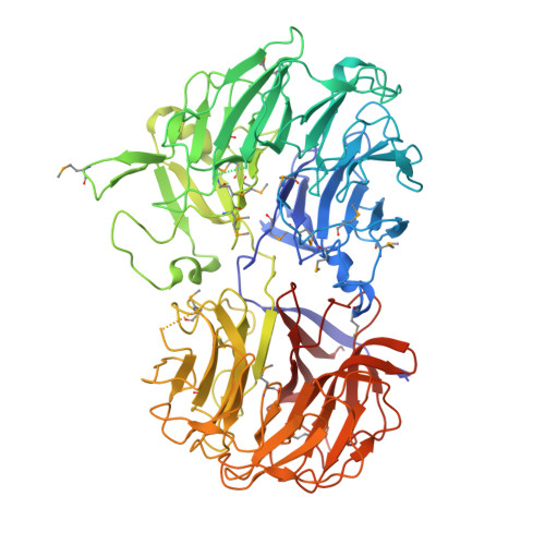
2CN2

Crystal Structures of Clostridium thermocellum Xyloglucanase


Experimental Data Snapshot

  • Method: X-RAY DIFFRACTION
  • Resolution: 2.10 Å
  • R-Value Free: 0.210 
  • R-Value Work: 0.176 
  • R-Value Observed: 0.178 

Starting Model: experimental
View more details

wwPDB Validation   3D Report Full Report


This is version 1.4 of the entry. See complete history


Literature

Crystal Structures of Clostridium Thermocellum Xyloglucanase, Xgh74A, Reveal the Structural Basis for Xyloglucan Recognition and Degradation.

Martinez-Fleites, C.Guerreiro, C.I.Baumann, M.J.Taylor, E.J.Prates, J.A.M.Ferreira, L.M.A.Fontes, C.M.G.A.Brumer, H.Davies, G.J.

(2006) J Biol Chem 281: 24922

  • DOI: https://doi.org/10.1074/jbc.M603583200
  • Primary Citation of Related Structures:  
    2CN2, 2CN3

  • PubMed Abstract: 

    The enzymatic degradation of the plant cell wall is central both to the natural carbon cycle and, increasingly, to environmentally friendly routes to biomass conversion, including the production of biofuels. The plant cell wall is a complex composite of cellulose microfibrils embedded in diverse polysaccharides collectively termed hemicelluloses. Xyloglucan is one such polysaccharide whose hydrolysis is catalyzed by diverse xyloglucanases. Here we present the structure of the Clostridium thermocellum xyloglucanase Xgh74A in both apo and ligand-complexed forms. The structures, in combination with mutagenesis data on the catalytic residues and the kinetics and specificity of xyloglucan hydrolysis reveal a complex subsite specificity accommodating seventeen monosaccharide moieties of the multibranched substrate in an open substrate binding terrain.


  • Organizational Affiliation

    York Structural Biology Laboratory, Department of Chemitry, University of York, York YO10 5YW, United Kingdom.


Macromolecules
Find similar proteins by:  (by identity cutoff)  |  3D Structure
Entity ID: 1
MoleculeChains Sequence LengthOrganismDetailsImage
BETA-1,4-XYLOGLUCAN HYDROLASE
A, B, C, D
737Acetivibrio thermocellusMutation(s): 0 
EC: 3.2.1.151 (PDB Primary Data), 3.2.1 (UniProt)
UniProt
Find proteins for Q70DK5 (Acetivibrio thermocellus)
Explore Q70DK5 
Go to UniProtKB:  Q70DK5
Entity Groups  
Sequence Clusters30% Identity50% Identity70% Identity90% Identity95% Identity100% Identity
UniProt GroupQ70DK5
Sequence Annotations
Expand
  • Reference Sequence
Small Molecules
Ligands 1 Unique
IDChains Name / Formula / InChI Key2D Diagram3D Interactions
CD
Query on CD

Download Ideal Coordinates CCD File 
E [auth A]
F [auth A]
G [auth A]
H [auth B]
I [auth B]
E [auth A],
F [auth A],
G [auth A],
H [auth B],
I [auth B],
J [auth B],
K [auth C],
L [auth C],
M [auth C],
N [auth C],
O [auth D],
P [auth D],
Q [auth D],
R [auth D],
S [auth D]
CADMIUM ION
Cd
WLZRMCYVCSSEQC-UHFFFAOYSA-N
Modified Residues  1 Unique
IDChains TypeFormula2D DiagramParent
MSE
Query on MSE
A, B, C, D
L-PEPTIDE LINKINGC5 H11 N O2 SeMET
Experimental Data & Validation

Experimental Data

  • Method: X-RAY DIFFRACTION
  • Resolution: 2.10 Å
  • R-Value Free: 0.210 
  • R-Value Work: 0.176 
  • R-Value Observed: 0.178 
  • Space Group: P 1 21 1
Unit Cell:
Length ( Å )Angle ( ˚ )
a = 100.437α = 90
b = 97.941β = 97.62
c = 199.122γ = 90
Software Package:
Software NamePurpose
REFMACrefinement
MOSFLMdata reduction
SCALAdata scaling
PHASERphasing

Structure Validation

View Full Validation Report


Entry History 

Revision History  (Full details and data files)

  • Version 1.0: 2006-05-22
    Type: Initial release
  • Version 1.1: 2011-05-08
    Changes: Version format compliance
  • Version 1.2: 2011-07-13
    Changes: Version format compliance
  • Version 1.3: 2023-12-13
    Changes: Data collection, Database references, Derived calculations, Other, Refinement description
  • Version 1.4: 2024-11-06
    Changes: Structure summary