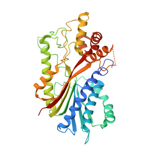
2EG5

The structure of xanthosine methyltransferase


Experimental Data Snapshot

  • Method: X-RAY DIFFRACTION
  • Resolution: 2.20 Å
  • R-Value Free: 0.284 
  • R-Value Work: 0.230 
  • R-Value Observed: 0.233 

Starting Model: experimental
View more details

wwPDB Validation   3D Report Full Report


Ligand Structure Quality Assessment 


This is version 1.3 of the entry. See complete history


Literature

The structure of two N-methyltransferases from the caffeine biosynthetic pathway

McCarthy, A.A.McCarthy, J.G.

(2007) Plant Physiol 144: 879-889

  • DOI: https://doi.org/10.1104/pp.106.094854
  • Primary Citation of Related Structures:  
    2EFJ, 2EG5

  • PubMed Abstract: 

    Caffeine (1,3,7-trimethylxanthine) is a secondary metabolite produced by certain plant species and an important component of coffee (Coffea arabica and Coffea canephora) and tea (Camellia sinensis). Here we describe the structures of two S-adenosyl-l-methionine-dependent N-methyltransferases that mediate caffeine biosynthesis in C. canephora 'robusta', xanthosine (XR) methyltransferase (XMT), and 1,7-dimethylxanthine methyltransferase (DXMT). Both were cocrystallized with the demethylated cofactor, S-adenosyl-L-cysteine, and substrate, either xanthosine or theobromine. Our structures reveal several elements that appear critical for substrate selectivity. Serine-316 in XMT appears central to the recognition of XR. Likewise, a change from glutamine-161 in XMT to histidine-160 in DXMT is likely to have catalytic consequences. A phenylalanine-266 to isoleucine-266 change in DXMT is also likely to be crucial for the discrimination between mono and dimethyl transferases in coffee. These key residues are probably functionally important and will guide future studies with implications for the biosynthesis of caffeine and its derivatives in plants.


  • Organizational Affiliation

    European Molecular Biology Laboratory, Grenoble 38042, France. andrewmc@embl.fr


Macromolecules
Find similar proteins by:  (by identity cutoff)  |  3D Structure
Entity ID: 1
MoleculeChains Sequence LengthOrganismDetailsImage
Xanthosine methyltransferaseA,
B [auth C],
C [auth E],
D [auth G]
372Coffea canephoraMutation(s): 0 
Gene Names: XMT1
EC: 2.1.1 (PDB Primary Data), 2.1.1.158 (UniProt)
UniProt
Find proteins for A4GE69 (Coffea canephora)
Explore A4GE69 
Go to UniProtKB:  A4GE69
Entity Groups  
Sequence Clusters30% Identity50% Identity70% Identity90% Identity95% Identity100% Identity
UniProt GroupA4GE69
Sequence Annotations
Expand
  • Reference Sequence
Small Molecules
Ligands 2 Unique
IDChains Name / Formula / InChI Key2D Diagram3D Interactions
SAH
Query on SAH

Download Ideal Coordinates CCD File 
E [auth A],
G [auth C],
I [auth E],
K [auth G]
S-ADENOSYL-L-HOMOCYSTEINE
C14 H20 N6 O5 S
ZJUKTBDSGOFHSH-WFMPWKQPSA-N
XTS
Query on XTS

Download Ideal Coordinates CCD File 
F [auth A],
H [auth C],
J [auth E],
L [auth G]
9-[(2R,3R,4S,5R)-3,4-DIHYDROXY-5-(HYDROXYMETHYL)OXOLAN-2-YL]-3H-PURINE-2,6-DIONE
C10 H12 N4 O6
UBORTCNDUKBEOP-BDXYJKHTSA-N
Experimental Data & Validation

Experimental Data

  • Method: X-RAY DIFFRACTION
  • Resolution: 2.20 Å
  • R-Value Free: 0.284 
  • R-Value Work: 0.230 
  • R-Value Observed: 0.233 
  • Space Group: P 1 21 1
Unit Cell:
Length ( Å )Angle ( ˚ )
a = 57.59α = 90
b = 119.8β = 102.16
c = 116.4γ = 90
Software Package:
Software NamePurpose
REFMACrefinement
MxCuBEdata collection
XDSdata reduction
XSCALEdata scaling
PHASERphasing

Structure Validation

View Full Validation Report


Ligand Structure Quality Assessment 


Entry History 

Deposition Data

Revision History  (Full details and data files)

  • Version 1.0: 2007-05-01
    Type: Initial release
  • Version 1.1: 2008-04-30
    Changes: Version format compliance
  • Version 1.2: 2011-07-13
    Changes: Advisory, Version format compliance
  • Version 1.3: 2023-10-25
    Changes: Data collection, Database references, Derived calculations, Refinement description, Structure summary