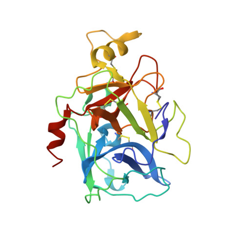
2F9O

Crystal Structure of the Recombinant Human Alpha I Tryptase Mutant D216G


Experimental Data Snapshot

  • Method: X-RAY DIFFRACTION
  • Resolution: 2.10 Å
  • R-Value Free: 0.245 
  • R-Value Work: 0.216 
  • R-Value Observed: 0.216 

Starting Model: experimental
View more details

wwPDB Validation   3D Report Full Report


This is version 2.3 of the entry. See complete history


Literature

X-ray Structures of Free and Leupeptin-complexed Human alpha I-Tryptase Mutants: Indication for an alpha to beta-Tryptase Transition

Rohr, K.B.Selwood, T.Marquardt, U.Huber, R.Schechter, N.M.Bode, W.Than, M.E.

(2005) J Mol Biol 357: 195-209

  • DOI: https://doi.org/10.1016/j.jmb.2005.12.037
  • Primary Citation of Related Structures:  
    2F9N, 2F9O, 2F9P

  • PubMed Abstract: 

    Tryptases alpha and beta are trypsin-like serine proteinases expressed in large amounts by mast cells. Beta-tryptase is a tetramer that has enzymatic activity, but requires heparin binding to maintain functional and structural stability, whereas alpha-tryptase has little, if any, enzymatic activity but is a stable tetramer in the absence of heparin. As shown previously, these differences can be mainly attributed to the different conformations of the 214-220 segment. Interestingly, the replacement of Asp216 by Gly, which is present in beta-tryptase, results in enzymatically active but less stable alpha-tryptase mutants. We have solved the crystal structures of both the single (D216G) and the double (K192Q/D216G) mutant forms of recombinant human alphaI-tryptase in complex with the peptide inhibitor leupeptin, as well as the structure of the non-inhibited single mutant. The inhibited mutants exhibited an open functional substrate binding site, while in the absence of an inhibitor, the open (beta-tryptase-like) and the closed (alpha-tryptase-like) conformations were present simultaneously. This shows that both forms are in a two-state equilibrium, which is influenced by the residues in the vicinity of the active site and by inhibitor/substrate binding. Novel insights regarding the observed stability differences as well as a potential proteolytic activity of wild-type alpha-tryptase, which may possess a cryptic active site, are discussed.


  • Organizational Affiliation

    Max-Planck-Institut für Biochemie, Abteilung Strukturforschung, Am Klopferspitz 18, 82152 Martinsried, Germany.


Macromolecules
Find similar proteins by:  (by identity cutoff)  |  3D Structure
Entity ID: 1
MoleculeChains Sequence LengthOrganismDetailsImage
Tryptase alpha-1
A, B, C, D
245Homo sapiensMutation(s): 2 
Gene Names: TPSAB1TPS1
EC: 3.4.21.59
UniProt & NIH Common Fund Data Resources
Find proteins for Q15661 (Homo sapiens)
Explore Q15661 
Go to UniProtKB:  Q15661
PHAROS:  Q15661
GTEx:  ENSG00000172236 
Entity Groups  
Sequence Clusters30% Identity50% Identity70% Identity90% Identity95% Identity100% Identity
UniProt GroupQ15661
Glycosylation
Glycosylation Sites: 1Go to GlyGen: Q15661-1
Sequence Annotations
Expand
  • Reference Sequence
Oligosaccharides

Help

Entity ID: 2
MoleculeChains Length2D Diagram Glycosylation3D Interactions
alpha-L-fucopyranose-(1-3)-[2-acetamido-2-deoxy-beta-D-glucopyranose-(1-4)]2-acetamido-2-deoxy-beta-D-glucopyranose
E, F
3N-Glycosylation
Glycosylation Resources
GlyTouCan:  G11971MR
GlyCosmos:  G11971MR
GlyGen:  G11971MR
Experimental Data & Validation

Experimental Data

  • Method: X-RAY DIFFRACTION
  • Resolution: 2.10 Å
  • R-Value Free: 0.245 
  • R-Value Work: 0.216 
  • R-Value Observed: 0.216 
  • Space Group: P 21 21 21
Unit Cell:
Length ( Å )Angle ( ˚ )
a = 83.31α = 90
b = 88.93β = 90
c = 162.92γ = 90
Software Package:
Software NamePurpose
DENZOdata reduction
SCALEPACKdata scaling
AMoREphasing
CNSrefinement

Structure Validation

View Full Validation Report


Entry History 

Deposition Data

Revision History  (Full details and data files)

  • Version 1.0: 2006-01-31
    Type: Initial release
  • Version 1.1: 2008-05-01
    Changes: Version format compliance
  • Version 1.2: 2011-07-13
    Changes: Non-polymer description, Version format compliance
  • Version 2.0: 2020-07-29
    Type: Remediation
    Reason: Carbohydrate remediation
    Changes: Advisory, Atomic model, Data collection, Derived calculations, Structure summary
  • Version 2.1: 2021-10-20
    Changes: Advisory, Database references, Structure summary
  • Version 2.2: 2023-08-30
    Changes: Data collection, Refinement description
  • Version 2.3: 2024-11-20
    Changes: Structure summary