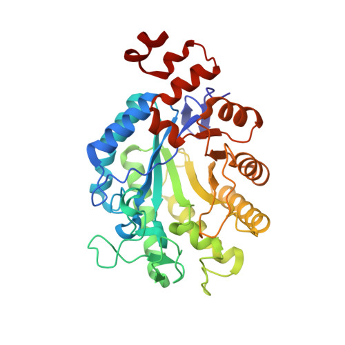
RCSB PDB - 2H8X: Xenobiotic Reductase A-oxidized

 2H8X

Xenobiotic Reductase A-oxidized


Experimental Data Snapshot

  • Method: X-RAY DIFFRACTION
  • Resolution: 1.50 Å
  • R-Value Free: 0.191 
  • R-Value Work: 0.182 
  • R-Value Observed: 0.184 

Starting Model: experimental
View more details

wwPDB Validation   3D Report Full Report


Ligand Structure Quality Assessment 

Created with Raphaël 2.3.0Worse 01 BetterLigand structure goodness of fit to experimental dataBest fitted FMNClick on this verticalbar to view details

This is version 1.4 of the entry. See complete history


Literature

Xenobiotic reductase A in the degradation of quinoline by Pseudomonas putida 86: physiological function, structure and mechanism of 8-hydroxycoumarin reduction.

Griese, J.J.Jakob, R.P.Schwarzinger, S.Dobbek, H.

(2006) J Mol Biol 361: 140-152

  • DOI: https://doi.org/10.1016/j.jmb.2006.06.007
  • Primary Citation of Related Structures:  
    2H8X, 2H8Z, 2H90

  • PubMed Abstract: 

    A continuous evolutionary pressure of the biotic and abiotic world has led to the development of a diversity of microbial pathways to degrade and biomineralize aromatic and heteroaromatic compounds. The heterogeneity of compounds metabolized by bacteria like Pseudomonas putida indicates the large variety of enzymes necessary to catalyse the required reactions. Quinoline, a N-heterocyclic aromatic compound, can be degraded by microbes along different pathways. For P. putida 86 quinoline degradation by the 8-hydroxycoumarin pathway has been described and several intermediates were identified. To select enzymes catalysing the later stages of the 8-hydroxycoumarin pathway P. putida 86 was grown with quinoline. The FMN-containing enzyme xenobiotic reductase A (XenA) was isolated and analysed for its reactivity with intermediates of the 8-hydroxycoumarin pathway. XenA catalyses the NADPH-dependent reduction of 8-hydroxycoumarin and coumarin to produce 8-hydroxy-3,4-dihydrocoumarin and 3,4-dihydrocoumarin, respectively. Crystallographic analysis of XenA alone and in complex with the two substrates revealed insights into the mechanism. XenA shows a dimeric arrangement of two (beta/alpha)(8) barrel domains each binding one FMN cofactor. High resolution crystal structures of complexes with 8-hydroxycoumarin and coumarin show different modes of binding for these molecules in the active site. While coumarin is ideally positioned for hydride transfer from N-5 of the isoalloxazine ring to C-4 of coumarin, 8-hydroxycoumarin forms a non-productive complex with oxidised XenA. Orientation of 8-hydroxycoumarin in the active site appears to be dependent on the electronic state of the flavin. We postulate that XenA catalyses the last step of the 8-hydroxycoumarin pathway before the heterocyclic ring is hydrolysed to yield 3-(2,3-dihydroxyphenyl)-propionic acid. As XenA is also found in P. putida strains unable to degrade quinoline, it appears to have more than one physiological function and is an example of how enzymes with low substrate specificity can help to explain the variety of degradation pathways possible.


  • Organizational Affiliation

    Laboratorium Proteinkristallographie, Universität Bayreuth, Germany.


Macromolecules
Find similar proteins by:  (by identity cutoff)  |  3D Structure
Entity ID: 1
MoleculeChains Sequence LengthOrganismDetailsImage
Xenobiotic reductase A358Pseudomonas putidaMutation(s): 0 
Gene Names: xenA
EC: 1.6.99.1
UniProt
Find proteins for Q88NF7 (Pseudomonas putida (strain ATCC 47054 / DSM 6125 / CFBP 8728 / NCIMB 11950 / KT2440))
Explore Q88NF7 
Go to UniProtKB:  Q88NF7
Entity Groups  
Sequence Clusters30% Identity50% Identity70% Identity90% Identity95% Identity100% Identity
UniProt GroupQ88NF7
Sequence Annotations
Expand
  • Reference Sequence
Small Molecules
Ligands 2 Unique
IDChains Name / Formula / InChI Key2D Diagram3D Interactions
FMN
Query on FMN

Download Ideal Coordinates CCD File 
K [auth A]FLAVIN MONONUCLEOTIDE
C17 H21 N4 O9 P
FVTCRASFADXXNN-SCRDCRAPSA-N
SO4
Query on SO4

Download Ideal Coordinates CCD File 
B [auth A]
C [auth A]
D [auth A]
E [auth A]
F [auth A]
SULFATE ION
O4 S
QAOWNCQODCNURD-UHFFFAOYSA-L
Experimental Data & Validation

Experimental Data

  • Method: X-RAY DIFFRACTION
  • Resolution: 1.50 Å
  • R-Value Free: 0.191 
  • R-Value Work: 0.182 
  • R-Value Observed: 0.184 
  • Space Group: I 2 2 2
Unit Cell:
Length ( Å )Angle ( ˚ )
a = 58.06α = 90
b = 83.35β = 90
c = 156.54γ = 90
Software Package:
Software NamePurpose
MAR345data collection
XDSdata reduction
SHARPphasing
CNSrefinement
XDSdata scaling

Structure Validation

View Full Validation Report


Ligand Structure Quality Assessment 

Created with Raphaël 2.3.0Worse 01 BetterLigand structure goodness of fit to experimental dataBest fitted FMNClick on this verticalbar to view details

Entry History 

Deposition Data

  • Released Date: 2006-08-29 
  • Deposition Author(s): Dobbek, H.

Revision History  (Full details and data files)

  • Version 1.0: 2006-08-29
    Type: Initial release
  • Version 1.1: 2008-05-01
    Changes: Version format compliance
  • Version 1.2: 2011-07-13
    Changes: Version format compliance
  • Version 1.3: 2017-10-18
    Changes: Refinement description
  • Version 1.4: 2023-08-30
    Changes: Data collection, Database references, Derived calculations, Refinement description