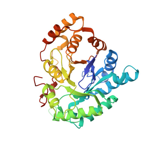
Crystal Structures of Mouse 17alpha-Hydroxysteroid Dehydrogenase (Apoenzyme and Enzyme-NADP(H) Binary Complex): Identification of Molecular Determinants Responsible for the Unique 17alpha-reductive Activity of this Enzyme.
Faucher, F., Pereira de Jesus-Tran, K., Cantin, L., Luu-The, V., Labrie, F., Breton, R.(2006) J Mol Biol 364: 747-763
- PubMed: 17034817
- DOI: https://doi.org/10.1016/j.jmb.2006.09.030
- Primary Citation of Related Structures:
2HDJ, 2HE5, 2HE8, 2HEJ - PubMed Abstract:
Very recently, the mouse 17alpha-hydroxysteroid dehydrogenase (m17alpha-HSD), a member of the aldo-keto reductase (AKR) superfamily, has been characterized and identified as the unique enzyme able to catalyze efficiently and in a stereospecific manner the conversion of androstenedione (Delta4) into epitestosterone (epi-T), the 17alpha-epimer of testosterone. Indeed, the other AKR enzymes that significantly reduce keto groups situated at position C17 of the steroid nucleus, the human type 3 3alpha-HSD (h3alpha-HSD3), the human and mouse type 5 17beta-HSD, and the rabbit 20alpha-HSD, produce only 17beta-hydroxy derivatives, although they possess more than 70% amino acid identity with m17alpha-HSD. Structural comparisons of these highly homologous enzymes thus offer an excellent opportunity of identifying the molecular determinants responsible for their 17alpha/17beta-stereospecificity. Here, we report the crystal structure of the m17alpha-HSD enzyme in its apo-form (1.9 A resolution) as well as those of two different forms of this enzyme in binary complex with NADP(H) (2.9 A and 1.35 A resolution). Interestingly, one of these binary complex structures could represent a conformational intermediate between the apoenzyme and the active binary complex. These structures provide a complete picture of the NADP(H)-enzyme interactions involving the flexible loop B, which can adopt two different conformations upon cofactor binding. Structural comparison with binary complexes of other AKR1C enzymes has also revealed particularities of the interaction between m17alpha-HSD and NADP(H), which explain why it has been possible to crystallize this enzyme in its apo form. Close inspection of the m17alpha-HSD steroid-binding cavity formed upon cofactor binding leads us to hypothesize that the residue at position 24 is of paramount importance for the stereospecificity of the reduction reaction. Mutagenic studies have showed that the m17alpha-HSD(A24Y) mutant exhibited a completely reversed stereospecificity, producing testosterone only from Delta4, whereas the h3alpha-HSD3(Y24A) mutant acquires the capacity to metabolize Delta4 into epi-T.
Organizational Affiliation:
Oncology and Molecular Endocrinology Research Center, Laval University Medical Center (CHUL) and Laval University, Québec (QC), Canada G1V 4G2.


