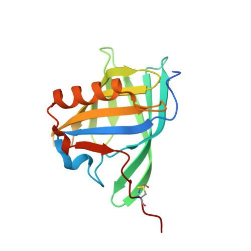
2HLV | pdb_00002hlv

Bovine Odorant Binding Protein deswapped triple mutant


Experimental Data Snapshot

  • Method: X-RAY DIFFRACTION
  • Resolution: 1.65 Å
  • R-Value Free: 
    0.212 (Depositor), 0.220 (DCC) 
  • R-Value Work: 
    0.185 (Depositor), 0.190 (DCC) 
  • R-Value Observed: 
    0.186 (Depositor) 

Starting Model: experimental
View more details

wwPDB Validation   3D Report Full Report


Ligand Structure Quality Assessment 

Created with Raphaël 2.3.0Worse 01 BetterLigand structure goodness of fit to experimental dataBest fitted LIKClick on this verticalbar to view details

This is version 1.4 of the entry. See complete history


Literature

Deswapping bovine odorant binding protein.

Ramoni, R.Spinelli, S.Grolli, S.Conti, V.Merli, E.Cambillau, C.Tegoni, M.

(2008) Biochim Biophys Acta 1784: 651-657

  • DOI: https://doi.org/10.1016/j.bbapap.2008.01.010
  • Primary Citation of Related Structures:  
    2HLV

  • PubMed Abstract: 

    The X-ray structure of bovine Odorant Binding Protein (bOBP) revealed its association as a domain swapped dimer. bOBP, devoid of any cysteines, contrasts with other mammalian OBPs, which are monomeric and possess at least one disulfide bridge. We have produced a mutant of bOBP in which a glycine residue was inserted after position 121. This mutation yielded a monomeric bOBP-121Gly+ in which domain swapping has been reverted. Here, we have subsequently introduced two mutations, Trp64Cys and His155Cys, in view to stabilize the putative monomer with a disulfide bridge. We have determined the crystal structure of this triple mutant at 1.65 A resolution. The mutant protein is monomeric, stabilized by a disulfide bridge between Trp64Cys and His155Cys, with a backbone superimposable to that of native bOBP, with the exception of the hinge and of the 10 residues at the C-terminus. bOBP triple mutant binds 1-amino-anthracene, 1-octen-3-ol (bOBP co-purified ligand) and other ligands with microM Kd values comparable to those of the swapped dimer.


  • Organizational Affiliation

    Dipartimento di Produzioni Animali, Biotecnologie Veterinarie, Qualità e Sicurezza degli Alimenti, Università degli Studi di Parma, Via del Taglio 8, 43100 Parma, Italy.


Macromolecules
Find similar proteins by:  (by identity cutoff)  |  3D Structure
Entity ID: 1
MoleculeChains Sequence LengthOrganismDetailsImage
Odorant-binding protein160Bos taurusMutation(s): 2 
UniProt
Find proteins for P07435 (Bos taurus)
Explore P07435 
Go to UniProtKB:  P07435
Entity Groups  
Sequence Clusters30% Identity50% Identity70% Identity90% Identity95% Identity100% Identity
UniProt GroupP07435
Sequence Annotations
Expand
  • Reference Sequence
Experimental Data & Validation

Experimental Data

  • Method: X-RAY DIFFRACTION
  • Resolution: 1.65 Å
  • R-Value Free:  0.212 (Depositor), 0.220 (DCC) 
  • R-Value Work:  0.185 (Depositor), 0.190 (DCC) 
  • R-Value Observed: 0.186 (Depositor) 
Space Group: P 41 21 2
Unit Cell:
Length ( Å )Angle ( ˚ )
a = 83.501α = 90
b = 83.501β = 90
c = 48.964γ = 90
Software Package:
Software NamePurpose
REFMACrefinement
DENZOdata reduction
SCALAdata scaling
AMoREphasing

Structure Validation

View Full Validation Report


Ligand Structure Quality Assessment 

Created with Raphaël 2.3.0Worse 01 BetterLigand structure goodness of fit to experimental dataBest fitted LIKClick on this verticalbar to view details

Entry History 

Deposition Data

Revision History  (Full details and data files)

  • Version 1.0: 2008-03-25
    Type: Initial release
  • Version 1.1: 2011-07-13
    Changes: Non-polymer description, Version format compliance
  • Version 1.2: 2021-10-20
    Changes: Database references, Derived calculations
  • Version 1.3: 2023-08-30
    Changes: Data collection, Refinement description
  • Version 1.4: 2024-10-09
    Changes: Structure summary