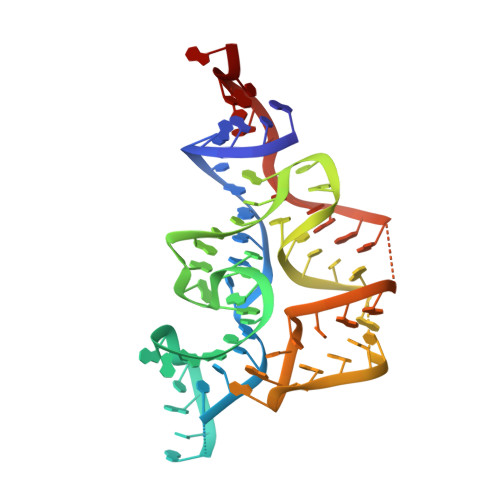
Crystal Structures of the Thi-Box Riboswitch Bound to Thiamine Pyrophosphate Analogs Reveal Adaptive RNA-Small Molecule Recognition
Edwards, T.E., Ferre-D'Amare, A.R.(2006) Structure 14: 1459-1468
- PubMed: 16962976
- DOI: https://doi.org/10.1016/j.str.2006.07.008
- Primary Citation of Related Structures:
2HOJ, 2HOK, 2HOL, 2HOM, 2HOO, 2HOP - PubMed Abstract:
Riboswitches are noncoding mRNA elements that bind small-molecule metabolites with high affinity and specificity, and they regulate the expression of associated genes. The thi-box riboswitch can exhibit a 1000-fold higher affinity for thiamine pyrophosphate over closely related noncognate compounds such as thiamine monophosphate. To understand the chemical basis of thi-box pyrophosphate specificity, we have determined crystal structures of an E. coli thi-box bound to thiamine pyrophosphate, thiamine monophosphate, and the structural analogs benfotiamine and pyrithiamine. When bound to monophosphorylated compounds, the RNA elements that recognize the thiamine and phosphate moieties of the ligand move closer together. This allows the riboswitch to recognize the monophosphate in a manner similar to how it recognizes the beta-phosphate of thiamine pyrophosphate. In the pyrithiamine complex, the pyrophosphate binding site is largely unstructured. These results show how the riboswitch can bind to various metabolites, and why the thi-box preferentially binds thiamine pyrophosphate.
Organizational Affiliation:
Division of Basic Sciences, Fred Hutchinson Cancer Research Center, Seattle, Washington 98109, USA.


