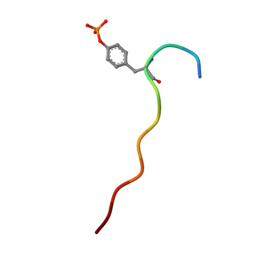
2IUI

Crystal structure of the PI3-kinase p85 N-terminal SH2 domain in complex with PDGFR phosphotyrosyl peptide


Experimental Data Snapshot

  • Method: X-RAY DIFFRACTION
  • Resolution: 2.40 Å
  • R-Value Free: 0.248 
  • R-Value Work: 0.178 
  • R-Value Observed: 0.178 

wwPDB Validation   3D Report Full Report


This is version 1.4 of the entry. See complete history


Literature

Crystal Structure of the Pi 3-Kinase P85 Amino- Terminal Sh2 Domain and its Phosphopeptide Complexes

Nolte, R.T.Eck, M.J.Schlessinger, J.Shoelson, S.E.Harrison, S.C.

(1996) Nat Struct Biol 3: 364

  • DOI: https://doi.org/10.1038/nsb0496-364
  • Primary Citation of Related Structures:  
    2IUG, 2IUH, 2IUI

  • PubMed Abstract: 

    Crystal structures of the amino-terminal SH2 domain of the p85alpha subunit of phosphatidylinositol (PI) 3-kinase, alone and in complex with phosphopeptides bearing pTyr-Met/Val-Xaa-Met motifs, show that phosphopeptides bind in the two-pronged manner seen in high-affinity Lck and Src SH2 complexes, with conserved interactions between the domain and the peptide segment from phosphotyrosine to Met+3. Peptide binding requires the rearrangement of a tyrosyl side chain in the BG loop to create the hydrophobic Met+3 binding pocket. The structures suggest a mechanism for the biological specificity exhibited by PI 3-kinase in its interactions with phosphoprotein partners.


  • Organizational Affiliation

    Howard Hughes Medical Institute and Laboratory of Molecular Medicine, Children's Hospital, Boston, Massachusetts 02115, USA.


Macromolecules
Find similar proteins by:  (by identity cutoff)  |  3D Structure
Entity ID: 1
MoleculeChains Sequence LengthOrganismDetailsImage
Phosphatidylinositol 3-kinase regulatory subunit alpha
A, B
120Homo sapiensMutation(s): 0 
Gene Names: PIK3R1GRB1
UniProt & NIH Common Fund Data Resources
Find proteins for P27986 (Homo sapiens)
Explore P27986 
Go to UniProtKB:  P27986
PHAROS:  P27986
GTEx:  ENSG00000145675 
Entity Groups  
Sequence Clusters30% Identity50% Identity70% Identity90% Identity95% Identity100% Identity
UniProt GroupP27986
Sequence Annotations
Expand
  • Reference Sequence

Find similar proteins by:  Sequence   |   3D Structure  

Entity ID: 2
MoleculeChains Sequence LengthOrganismDetailsImage
Platelet-derived growth factor receptor beta
C, D
11Homo sapiensMutation(s): 0 
EC: 2.7.10.1
UniProt & NIH Common Fund Data Resources
Find proteins for P09619 (Homo sapiens)
Explore P09619 
Go to UniProtKB:  P09619
PHAROS:  P09619
GTEx:  ENSG00000113721 
Entity Groups  
Sequence Clusters30% Identity50% Identity70% Identity90% Identity95% Identity100% Identity
UniProt GroupP09619
Sequence Annotations
Expand
  • Reference Sequence
Small Molecules
Modified Residues  1 Unique
IDChains TypeFormula2D DiagramParent
PTR
Query on PTR
C, D
L-PEPTIDE LINKINGC9 H12 N O6 PTYR
Experimental Data & Validation

Experimental Data

  • Method: X-RAY DIFFRACTION
  • Resolution: 2.40 Å
  • R-Value Free: 0.248 
  • R-Value Work: 0.178 
  • R-Value Observed: 0.178 
  • Space Group: P 21 21 21
Unit Cell:
Length ( Å )Angle ( ˚ )
a = 40.76α = 90
b = 61.94β = 90
c = 108.36γ = 90
Software Package:
Software NamePurpose
X-PLORrefinement

Structure Validation

View Full Validation Report


Entry History 

Deposition Data

Revision History  (Full details and data files)

  • Version 1.0: 2006-06-06
    Type: Initial release
  • Version 1.1: 2011-05-08
    Changes: Version format compliance
  • Version 1.2: 2011-07-13
    Changes: Version format compliance
  • Version 1.3: 2018-03-07
    Changes: Database references, Source and taxonomy, Structure summary
  • Version 1.4: 2021-04-28
    Changes: Data collection, Derived calculations, Refinement description