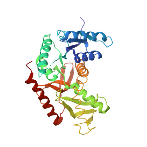
2LDB

STRUCTURE DETERMINATION AND REFINEMENT OF BACILLUS STEAROTHERMOPHILUS LACTATE DEHYDROGENASE


Experimental Data Snapshot

  • Method: X-RAY DIFFRACTION
  • Resolution: 3.00 Å
  • R-Value Observed: 0.260 

wwPDB Validation   3D Report Full Report


This is version 2.1 of the entry. See complete history


Literature

Structure determination and refinement of Bacillus stearothermophilus lactate dehydrogenase.

Piontek, K.Chakrabarti, P.Schar, H.P.Rossmann, M.G.Zuber, H.

(1990) Proteins 7: 74-92

  • DOI: https://doi.org/10.1002/prot.340070108
  • Primary Citation of Related Structures:  
    1LDB, 2LDB

  • PubMed Abstract: 

    Structures have been determined of Bacillus stearothermophilus "apo" and holo lactate dehydrogenase. The holo-enzyme had been co-crystallized with the activator fructose 1,6-bisphosphate. The "apo" lactate dehydrogenase structure was solved by use of the known apo-M4 dogfish lactate dehydrogenase molecule as a starting model. Phases were refined and extended from 4 A to 3 A resolution by means of the noncrystallographic molecular 222 symmetry. The R-factor was reduced to 28.7%, using 2.8 A resolution data, in a restrained least-squares refinement in which the molecular symmetry was imposed as a constraint. A low occupancy of coenzyme was found in each of the four subunits of the "apo"-enzyme. Further refinement proceeded with the isomorphous holo-enzyme from Bacillus stearothermophilus. After removing the noncrystallographic constraints, the R-factor dropped from 30.3% to a final value of 26.0% with a 0.019 A and 1.7 degrees r.m.s. deviation from idealized bond lengths and angles, respectively. Two sulfate ions per subunit were included in the final model of the "apo"-form--one at the substrate binding site and one close to the molecular P-axis near the location of the fructose 1,6-bisphosphate activator. The final model of the holo-enzyme incorporated two sulfate ions per subunit, one at the substrate binding site and another close to the R-axis. One nicotinamide adenine dinucleotide coenzyme molecule per subunit and two fructose 1,6-bisphosphate molecules per tetramer were also included. The phosphate positions of fructose 1,6-bisphosphate are close to the sulfate ion near the P-axis in the "apo" model. This structure represents the first reported refined model of an allosteric activated lactate dehydrogenase. The structure of the activated holo-enzyme showed far greater similarity to the ternary complex of dogfish M4 lactate dehydrogenase with nicotinamide adenine dinucleotide and oxamate than to apo-M4 dogfish lactate dehydrogenase. The conformations of nicotinamide adenine dinucleotide and fructose 1,6-bisphosphate were also analyzed.


  • Organizational Affiliation

    Department of Biological Sciences, Purdue University, West Lafayette, Indiana 47907.


Macromolecules
Find similar proteins by:  (by identity cutoff)  |  3D Structure
Entity ID: 1
MoleculeChains Sequence LengthOrganismDetailsImage
L-LACTATE DEHYDROGENASE
A, B, C, D
317Geobacillus stearothermophilusMutation(s): 0 
EC: 1.1.1.27
UniProt
Find proteins for P00344 (Geobacillus stearothermophilus)
Explore P00344 
Go to UniProtKB:  P00344
Entity Groups  
Sequence Clusters30% Identity50% Identity70% Identity90% Identity95% Identity100% Identity
UniProt GroupP00344
Sequence Annotations
Expand
  • Reference Sequence
Small Molecules
Ligands 3 Unique
IDChains Name / Formula / InChI Key2D Diagram3D Interactions
NAD
Query on NAD

Download Ideal Coordinates CCD File 
G [auth A],
J [auth B],
N [auth C],
R [auth D]
NICOTINAMIDE-ADENINE-DINUCLEOTIDE
C21 H27 N7 O14 P2
BAWFJGJZGIEFAR-NNYOXOHSSA-N
FBP
Query on FBP

Download Ideal Coordinates CCD File 
K [auth C],
O [auth D]
1,6-di-O-phosphono-beta-D-fructofuranose
C6 H14 O12 P2
RNBGYGVWRKECFJ-ARQDHWQXSA-N
SO4
Query on SO4

Download Ideal Coordinates CCD File 
E [auth A]
F [auth A]
H [auth B]
I [auth B]
L [auth C]
E [auth A],
F [auth A],
H [auth B],
I [auth B],
L [auth C],
M [auth C],
P [auth D],
Q [auth D]
SULFATE ION
O4 S
QAOWNCQODCNURD-UHFFFAOYSA-L
Experimental Data & Validation

Experimental Data

  • Method: X-RAY DIFFRACTION
  • Resolution: 3.00 Å
  • R-Value Observed: 0.260 
  • Space Group: P 61
Unit Cell:
Length ( Å )Angle ( ˚ )
a = 86.8α = 90
b = 86.8β = 90
c = 356.6γ = 120
Software Package:
Software NamePurpose
PROLSQrefinement

Structure Validation

View Full Validation Report


Entry History 

Deposition Data

Revision History  (Full details and data files)

  • Version 1.0: 1989-07-12
    Type: Initial release
  • Version 1.1: 2008-03-03
    Changes: Version format compliance
  • Version 1.2: 2011-07-13
    Changes: Version format compliance
  • Version 1.3: 2020-07-29
    Type: Remediation
    Reason: Carbohydrate remediation
    Changes: Data collection, Derived calculations, Other, Structure summary
  • Version 2.0: 2023-09-27
    Type: Remediation
    Changes: Advisory, Atomic model, Data collection, Database references, Other, Refinement description, Structure summary
  • Version 2.1: 2024-12-25
    Changes: Advisory, Derived calculations, Structure summary