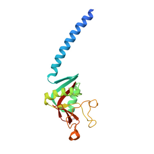
2ORJ

crystal structure of the trimeric neck and carbohydrate recognition domain of human surfactant protein D in complex with N-acetyl mannosamine


Experimental Data Snapshot

  • Method: X-RAY DIFFRACTION
  • Resolution: 1.80 Å
  • R-Value Free: 0.205 
  • R-Value Work: 0.178 
  • R-Value Observed: 0.181 

Starting Model: experimental
View more details

wwPDB Validation   3D Report Full Report


Ligand Structure Quality Assessment 


This is version 1.5 of the entry. See complete history


Literature

Critical Role of Arg/Lys343 in the Species-Dependent Recognition of Phosphatidylinositol by Pulmonary Surfactant Protein D.

Crouch, E.McDonald, B.Smith, K.Roberts, M.Mealy, T.Seaton, B.Head, J.

(2007) Biochemistry 46: 5160-5169

  • DOI: https://doi.org/10.1021/bi700037x
  • Primary Citation of Related Structures:  
    2ORJ, 2ORK, 2OS9

  • PubMed Abstract: 

    Surfactant protein D (SP-D) plays important roles in lung host defense. However, it can also recognize specific host molecules and contributes to surfactant homeostasis. The major known surfactant-associated ligand is phosphatidylinositol (PI). Trimeric neck-carbohydrate recognition domains (NCRDs) of rat and human SP-D exhibited dose-dependent, calcium-dependent, and inositol-sensitive binding to solid-phase PI and to multilamellar PI liposomes. However, the rat protein exhibited a >5-fold higher affinity for solid-phase PI than the human NCRD. In addition, human dodecamers, but not full-length human trimers, efficiently coprecipitated with multilamellar PI liposomes in the presence of calcium. A human NCRD mutant resembling the rat and mouse proteins at position 343 (hR343K) showed much stronger binding to PI. A reciprocal rat mutant with arginine at the position of lysine 343 (rK343R) showed weak binding to PI, even weaker than that of the wild-type human protein. Crystal complexes of the human trimeric NCRD with myoinositol and inositol 1-phosphate showed binding of the equatorial OH groups of the cyclitol ring of the inositol to calcium at the carbohydrate binding site. Myoinositol binding occurred in two major orientations, while inositol 1-phosphate appeared primarily constrained to a single, different orientation. Our studies directly implicate the CRD in PI binding and reveal unexpected species differences in PI recognition that can be largely attributed to the side chain of residue 343. In addition, the studies indicate that oligomerization of trimeric subunits is an important determinant of recognition of PI by human SP-D.


  • Organizational Affiliation

    Department of Pathology and Immunology, Washington University School of Medicine, St. Louis, Missouri 63110, USA. crouch@path.wustl.edu


Macromolecules
Find similar proteins by:  (by identity cutoff)  |  3D Structure
Entity ID: 1
MoleculeChains Sequence LengthOrganismDetailsImage
Pulmonary surfactant-associated protein D
A, B, C
160Homo sapiensMutation(s): 0 
Gene Names: SFTPDPSPDSFTP4
UniProt & NIH Common Fund Data Resources
Find proteins for P35247 (Homo sapiens)
Explore P35247 
Go to UniProtKB:  P35247
PHAROS:  P35247
GTEx:  ENSG00000133661 
Entity Groups  
Sequence Clusters30% Identity50% Identity70% Identity90% Identity95% Identity100% Identity
UniProt GroupP35247
Sequence Annotations
Expand
  • Reference Sequence
Small Molecules
Ligands 2 Unique
IDChains Name / Formula / InChI Key2D Diagram3D Interactions
BM3
Query on BM3

Download Ideal Coordinates CCD File 
G [auth A],
K [auth B],
O [auth C]
2-acetamido-2-deoxy-alpha-D-mannopyranose
C8 H15 N O6
OVRNDRQMDRJTHS-UOLFYFMNSA-N
CA
Query on CA

Download Ideal Coordinates CCD File 
D [auth A]
E [auth A]
F [auth A]
H [auth B]
I [auth B]
D [auth A],
E [auth A],
F [auth A],
H [auth B],
I [auth B],
J [auth B],
L [auth C],
M [auth C],
N [auth C]
CALCIUM ION
Ca
BHPQYMZQTOCNFJ-UHFFFAOYSA-N
Experimental Data & Validation

Experimental Data

  • Method: X-RAY DIFFRACTION
  • Resolution: 1.80 Å
  • R-Value Free: 0.205 
  • R-Value Work: 0.178 
  • R-Value Observed: 0.181 
  • Space Group: P 1 21 1
Unit Cell:
Length ( Å )Angle ( ˚ )
a = 55.63α = 90
b = 108.33β = 91.51
c = 55.97γ = 90
Software Package:
Software NamePurpose
REFMACrefinement
HKL-2000data collection
HKL-2000data reduction
HKL-2000data scaling
CNSphasing

Structure Validation

View Full Validation Report


Ligand Structure Quality Assessment 


Entry History 

Deposition Data

  • Released Date: 2007-05-08 
  • Deposition Author(s): Head, J.F.

Revision History  (Full details and data files)

  • Version 1.0: 2007-05-08
    Type: Initial release
  • Version 1.1: 2008-05-01
    Changes: Version format compliance
  • Version 1.2: 2011-07-13
    Changes: Version format compliance
  • Version 1.3: 2020-07-29
    Type: Remediation
    Reason: Carbohydrate remediation
    Changes: Advisory, Data collection, Database references, Derived calculations, Structure summary
  • Version 1.4: 2023-08-30
    Changes: Data collection, Database references, Refinement description, Structure summary
  • Version 1.5: 2024-10-30
    Changes: Structure summary