2PUQ

Crystal structure of active site inhibited coagulation factor VIIA in complex with soluble tissue factor


Experimental Data Snapshot

  • Method: X-RAY DIFFRACTION
  • Resolution: 2.05 Å
  • R-Value Free: 0.273 
  • R-Value Work: 0.228 
  • R-Value Observed: 0.231 

Starting Model: experimental
View more details

wwPDB Validation   3D Report Full Report


Ligand Structure Quality Assessment 


This is version 1.6 of the entry. See complete history


Literature

Engineering the substrate and inhibitor specificities of human coagulation Factor VIIa

Larsen, K.S.Ostergaard, H.Bjelke, J.R.Olsen, O.H.Rasmussen, H.B.Christensen, L.Kragelund, B.B.Stennicke, H.R.

(2007) Biochem J 405: 429-438

  • DOI: https://doi.org/10.1042/BJ20061901
  • Primary Citation of Related Structures:  
    2PUQ

  • PubMed Abstract: 

    The remarkably high specificity of the coagulation proteases towards macromolecular substrates is provided by numerous interactions involving the catalytic groove and remote exosites. For FVIIa [activated FVII (Factor VII)], the principal initiator of coagulation via the extrinsic pathway, several exosites have been identified, whereas only little is known about the specificity dictated by the active-site architecture. In the present study, we have profiled the primary P4-P1 substrate specificity of FVIIa using positional scanning substrate combinatorial libraries and evaluated the role of the selective active site in defining specificity. Being a trypsin-like serine protease, FVIIa had P1 specificity exclusively towards arginine and lysine residues. In the S2 pocket, threonine, leucine, phenylalanine and valine residues were the most preferred amino acids. Both S3 and S4 appeared to be rather promiscuous, however, with some preference for aromatic amino acids at both positions. Interestingly, a significant degree of interdependence between the S3 and S4 was observed and, as a consequence, the optimal substrate for FVIIa could not be derived directly from a subsite-directed specificity screen. To evaluate the role of the active-site residues in defining specificity, a series of mutants of FVIIa were prepared at position 239 (position 99 in chymotrypsin), which is considered to be one of the most important residues for determining P2 specificity of the trypsin family members. This was confirmed for FVIIa by marked changes in primary substrate specificity and decreased rates of antithrombin III inhibition. Interestingly, these changes do not necessarily coincide with an altered ability to activate Factor X, demonstrating that inhibitor and macromolecular substrate selectivity may be engineered separately.


  • Organizational Affiliation

    Haemostasis Biochemistry, Novo Nordisk A/S, Novo Nordisk Park, DK-2760 Måløv, Denmark. ksln@novonordisk.com


Macromolecules
Find similar proteins by:  (by identity cutoff)  |  3D Structure
Entity ID: 1
MoleculeChains Sequence LengthOrganismDetailsImage
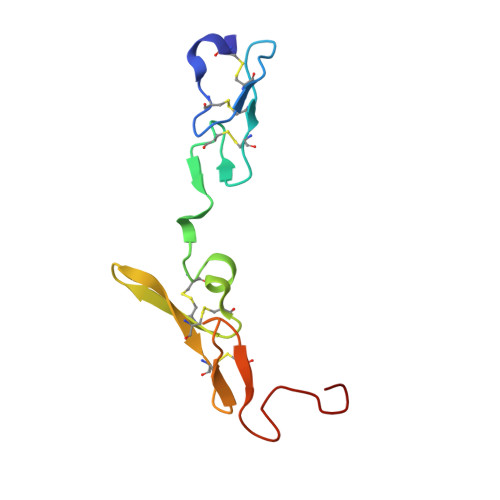
Coagulation factor VIIA [auth L]94Homo sapiensMutation(s): 0 
Gene Names: F7
EC: 3.4.21.21
UniProt & NIH Common Fund Data Resources
Find proteins for P08709 (Homo sapiens)
Explore P08709 
Go to UniProtKB:  P08709
PHAROS:  P08709
GTEx:  ENSG00000057593 
Entity Groups  
Sequence Clusters30% Identity50% Identity70% Identity90% Identity95% Identity100% Identity
UniProt GroupP08709
Glycosylation
Glycosylation Sites: 2Go to GlyGen: P08709-1
Sequence Annotations
Expand
  • Reference Sequence
Find similar proteins by:  (by identity cutoff)  |  3D Structure
Entity ID: 2
MoleculeChains Sequence LengthOrganismDetailsImage
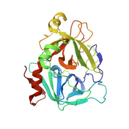
Coagulation factor VIIB [auth H]254Homo sapiensMutation(s): 0 
Gene Names: F7
EC: 3.4.21.21
UniProt & NIH Common Fund Data Resources
Find proteins for P08709 (Homo sapiens)
Explore P08709 
Go to UniProtKB:  P08709
PHAROS:  P08709
GTEx:  ENSG00000057593 
Entity Groups  
Sequence Clusters30% Identity50% Identity70% Identity90% Identity95% Identity100% Identity
UniProt GroupP08709
Sequence Annotations
Expand
  • Reference Sequence
Find similar proteins by:  (by identity cutoff)  |  3D Structure
Entity ID: 3
MoleculeChains Sequence LengthOrganismDetailsImage
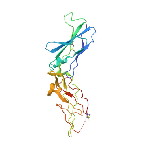
Tissue factorC [auth T]204Homo sapiensMutation(s): 0 
Gene Names: ACE
UniProt & NIH Common Fund Data Resources
Find proteins for P13726 (Homo sapiens)
Explore P13726 
Go to UniProtKB:  P13726
PHAROS:  P13726
GTEx:  ENSG00000117525 
Entity Groups  
Sequence Clusters30% Identity50% Identity70% Identity90% Identity95% Identity100% Identity
UniProt GroupP13726
Sequence Annotations
Expand
  • Reference Sequence

Find similar proteins by:  Sequence   |   3D Structure  

Entity ID: 4
MoleculeChains Sequence LengthOrganismDetailsImage
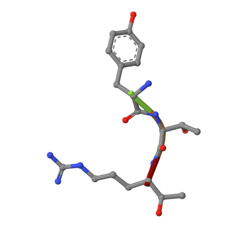
TRP-TYR-THR-ARG CHLOROMETHYLKETONE INHIBITORD [auth I]5N/AMutation(s): 0 
Sequence Annotations
Expand
  • Reference Sequence
Small Molecules
Experimental Data & Validation

Experimental Data

  • Method: X-RAY DIFFRACTION
  • Resolution: 2.05 Å
  • R-Value Free: 0.273 
  • R-Value Work: 0.228 
  • R-Value Observed: 0.231 
  • Space Group: P 1 21 1
Unit Cell:
Length ( Å )Angle ( ˚ )
a = 78.312α = 90
b = 68.827β = 90.74
c = 78.732γ = 90
Software Package:
Software NamePurpose
REFMACrefinement
PDB_EXTRACTdata extraction
XDSdata scaling
XDSdata reduction
PHASERphasing

Structure Validation

View Full Validation Report


Ligand Structure Quality Assessment 


Entry History 

Deposition Data

Revision History  (Full details and data files)

  • Version 1.0: 2007-05-22
    Type: Initial release
  • Version 1.1: 2008-05-01
    Changes: Version format compliance
  • Version 1.2: 2011-07-13
    Changes: Atomic model, Database references, Derived calculations, Non-polymer description, Structure summary, Version format compliance
  • Version 1.3: 2012-12-12
    Changes: Other
  • Version 1.4: 2020-07-29
    Type: Remediation
    Reason: Carbohydrate remediation
    Changes: Advisory, Data collection, Derived calculations, Structure summary
  • Version 1.5: 2023-08-30
    Changes: Advisory, Data collection, Database references, Refinement description, Structure summary
  • Version 1.6: 2024-11-13
    Changes: Structure summary