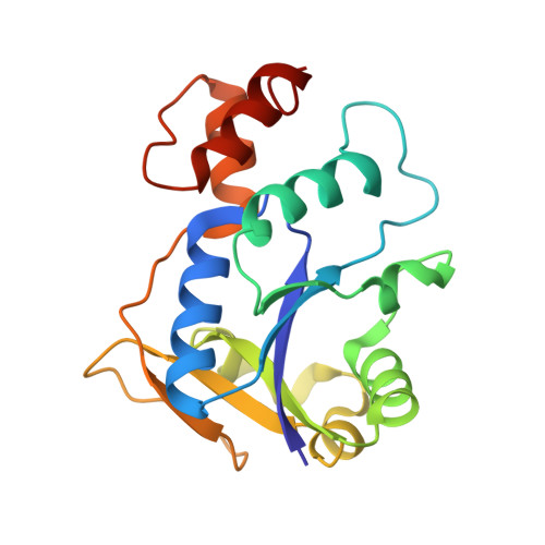
2QTN

Crystal Structure of Nicotinate Mononucleotide Adenylyltransferase


Experimental Data Snapshot

  • Method: X-RAY DIFFRACTION
  • Resolution: 2.40 Å
  • R-Value Free: 0.287 
  • R-Value Work: 0.204 

Starting Model: experimental
View more details

wwPDB Validation   3D Report Full Report


Ligand Structure Quality Assessment 


This is version 1.3 of the entry. See complete history


Literature

Kinetic and X-ray structural evidence for negative cooperativity in substrate binding to nicotinate mononucleotide adenylyltransferase (NMAT) from Bacillus anthracis.

Sershon, V.C.Santarsiero, B.D.Mesecar, A.D.

(2009) J Mol Biol 385: 867-888

  • DOI: https://doi.org/10.1016/j.jmb.2008.10.037
  • Primary Citation of Related Structures:  
    2QTM, 2QTN, 2QTR

  • PubMed Abstract: 

    Biosynthesis of NAD(P) in bacteria occurs either de novo or through one of the salvage pathways that converge at the point where the reaction of nicotinate mononucleotide (NaMN) with ATP is coupled to the formation of nicotinate adenine dinucleotide (NaAD) and inorganic pyrophosphate. This reaction is catalyzed by nicotinate mononucleotide adenylyltransferase (NMAT), which is essential for bacterial growth, making it an attractive drug target for the development of new antibiotics. Steady-state kinetic and direct binding studies on NMAT from Bacillus anthracis suggest a random sequential Bi-Bi kinetic mechanism. Interestingly, the interactions of NaMN and ATP with NMAT were observed to exhibit negative cooperativity, i.e. Hill coefficients <1.0. Negative cooperativity in binding is supported by the results of X-ray crystallographic studies. X-ray structures of the B. anthracis NMAT apoenzyme, and the NaMN- and NaAD-bound complexes were determined to resolutions of 2.50 A, 2.60 A and 1.75 A, respectively. The X-ray structure of the NMAT-NaMN complex revealed only one NaMN molecule bound in the biological dimer, supporting negative cooperativity in substrate binding. The kinetic, direct-binding, and X-ray structural studies support a model in which the binding affinity of substrates to the first monomer of NMAT is stronger than that to the second, and analysis of the three X-ray structures reveals significant conformational changes of NMAT along the enzymatic reaction coordinate. The negative cooperativity observed in B. anthracis NMAT substrate binding is a unique property that has not been observed in other prokaryotic NMAT enzymes. We propose that regulation of the NAD(P) biosynthetic pathway may occur, in part, at the reaction catalyzed by NMAT.


  • Organizational Affiliation

    Department of Medicinal Chemistry and Pharmacognosy & the Center for Pharmaceutical Biotechnology, University of Illinois at Chicago, Chicago, IL 60607, USA.


Macromolecules
Find similar proteins by:  (by identity cutoff)  |  3D Structure
Entity ID: 1
MoleculeChains Sequence LengthOrganismDetailsImage
Nicotinate (Nicotinamide) nucleotide adenylyltransferase
A, B
189Bacillus anthracisMutation(s): 0 
Gene Names: NadD
EC: 2.7.7.18
Entity Groups  
Sequence Clusters30% Identity50% Identity70% Identity90% Identity95% Identity100% Identity
Sequence Annotations
Expand
  • Reference Sequence
Small Molecules
Ligands 3 Unique
IDChains Name / Formula / InChI Key2D Diagram3D Interactions
NCN
Query on NCN

Download Ideal Coordinates CCD File 
H [auth B]NICOTINATE MONONUCLEOTIDE
C11 H14 N O9 P
JOUIQRNQJGXQDC-ZYUZMQFOSA-N
GOL
Query on GOL

Download Ideal Coordinates CCD File 
D [auth A]
E [auth A]
F [auth A]
I [auth B]
J [auth B]
D [auth A],
E [auth A],
F [auth A],
I [auth B],
J [auth B],
K [auth B]
GLYCEROL
C3 H8 O3
PEDCQBHIVMGVHV-UHFFFAOYSA-N
MG
Query on MG

Download Ideal Coordinates CCD File 
C [auth A],
G [auth B]
MAGNESIUM ION
Mg
JLVVSXFLKOJNIY-UHFFFAOYSA-N
Binding Affinity Annotations 
IDSourceBinding Affinity
NCN PDBBind:  2QTN Kd: 3.82e+5 (nM) from 1 assay(s)
Experimental Data & Validation

Experimental Data

  • Method: X-RAY DIFFRACTION
  • Resolution: 2.40 Å
  • R-Value Free: 0.287 
  • R-Value Work: 0.204 
  • Space Group: P 61
Unit Cell:
Length ( Å )Angle ( ˚ )
a = 95.895α = 90
b = 95.895β = 90
c = 84.532γ = 120
Software Package:
Software NamePurpose
DENZOdata reduction
SCALEPACKdata scaling
CNSrefinement
PDB_EXTRACTdata extraction
HKL-2000data collection
HKL-2000data reduction
HKL-2000data scaling
PHASERphasing

Structure Validation

View Full Validation Report


Ligand Structure Quality Assessment 


Entry History 

Deposition Data

Revision History  (Full details and data files)

  • Version 1.0: 2008-07-15
    Type: Initial release
  • Version 1.1: 2011-07-13
    Changes: Non-polymer description, Version format compliance
  • Version 1.2: 2017-10-25
    Changes: Refinement description
  • Version 1.3: 2023-08-30
    Changes: Data collection, Database references, Derived calculations, Refinement description