Structural and Mechanistic Studies of Arylalkylhydrazine Inhibition of Human Monoamine Oxidases a and B
Binda, C., Wang, J., Li, M., Hubalek, F., Mattevi, A., Edmondson, D.E.(2008) Biochemistry 47: 5616
- PubMed: 18426226
- DOI: https://doi.org/10.1021/bi8002814
- Primary Citation of Related Structures:
2VRL, 2VRM - PubMed Abstract:
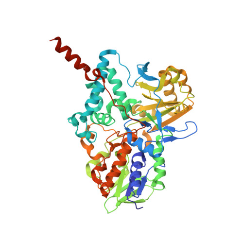
The structure and mechanism of human monoamine oxidase B (MAO B) inhibition by hydrazines are investigated and compared with data on human monoamine oxidase A (MAO A). The inhibition properties of phenylethylhydrazine, benzylhydrazine, and phenylhydrazine are compared for both enzymes. Benzylhydrazine is bound more tightly to MAO B than to MAO A, and phenylhydrazine is bound weakly by either enzyme. Phenylethylhydrazine stoichiometrically reduces the covalent FAD moieties of MAO A and of MAO B. Molecular oxygen is required for the inhibition reactions, and the level of O2 consumption for phenylethylhydrazine is 6-7-fold higher with either MAO A or MAO B than for the corresponding reactions with benzylhydrazine or phenylhydrazine. Mass spectral analysis of either inhibited enzyme shows the major product is a single covalent addition of the hydrazine arylalkyl group, although lower levels of dialkylated species are detected. Absorption and mass spectral data of the inhibited enzymes show that the FAD is the major site of alkylation. The three-dimensional (2.3 A) structures of phenylethylhydrazine- and benzylhydrazine-inhibited MAO B show that alkylation occurs at the N(5) position on the re face of the covalent flavin with loss of the hydrazyl nitrogens. A mechanistic scheme is proposed to account for these data, which involves enzyme-catalyzed conversion of the hydrazine to the diazene. From literature data on the reactivities of diazenes, O2 then reacts with the bound diazene to form an alkyl radical, N2 and superoxide anion. The bound arylalkyl radical reacts with the N(5) of the flavin, while the dissociated diazene reacts nonspecifically with the enzyme through arylalkylradicals.
Organizational Affiliation:
Department of Genetics and Microbiology, University of Pavia, via Ferrata 1, Pavia 27100, Italy.


