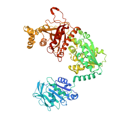
2VXO

Human GMP synthetase in complex with XMP


Experimental Data Snapshot

  • Method: X-RAY DIFFRACTION
  • Resolution: 2.50 Å
  • R-Value Free: 0.256 
  • R-Value Work: 0.203 
  • R-Value Observed: 0.206 

Starting Model: experimental
View more details

wwPDB Validation   3D Report Full Report


Ligand Structure Quality Assessment 


This is version 1.4 of the entry. See complete history


Literature

Substrate Specificity and Oligomerization of Human Gmp Synthetase

Welin, M.Lehtio, L.Johansson, A.Flodin, S.Nyman, T.Tresaugues, L.Hammarstrom, M.Graslund, S.Nordlund, P.

(2013) J Mol Biol 425: 4323

  • DOI: https://doi.org/10.1016/j.jmb.2013.06.032
  • Primary Citation of Related Structures:  
    2VXO

  • PubMed Abstract: 

    Guanine monophosphate (GMP) synthetase is a bifunctional two-domain enzyme. The N-terminal glutaminase domain generates ammonia from glutamine and the C-terminal synthetase domain aminates xanthine monophosphate (XMP) to form GMP. Mammalian GMP synthetases (GMPSs) contain a 130-residue-long insert in the synthetase domain in comparison to bacterial proteins. We report here the structure of a eukaryotic GMPS. Substrate XMP was bound in the crystal structure of the human GMPS enzyme. XMP is bound to the synthetase domain and covered by a LID motif. The enzyme forms a dimer in the crystal structure with subunit orientations entirely different from the bacterial counterparts. The inserted sub-domain is shown to be involved in substrate binding and dimerization. Furthermore, the structural basis for XMP recognition is revealed as well as a potential allosteric site. Enzymes in the nucleotide metabolism typically display an increased activity in proliferating cells due to the increased need for nucleotides. Many drugs used as immunosuppressants and for treatment of cancer and viral diseases are indeed nucleobase- and nucleoside-based compounds, which are acting on or are activated by enzymes in this pathway. The information obtained from the crystal structure of human GMPS might therefore aid in understanding interactions of nucleoside-based drugs with GMPS and in structure-based design of GMPS-specific inhibitors.


  • Organizational Affiliation

    Structural Genomics Consortium, Department of Medical Biochemistry and Biophysics, Karolinska Institutet, S-17177 Stockholm, Sweden.


Macromolecules
Find similar proteins by:  (by identity cutoff)  |  3D Structure
Entity ID: 1
MoleculeChains Sequence LengthOrganismDetailsImage
GMP SYNTHASE [GLUTAMINE-HYDROLYZING]
A, B
697Homo sapiensMutation(s): 0 
EC: 6.3.5.2
UniProt & NIH Common Fund Data Resources
Find proteins for P49915 (Homo sapiens)
Explore P49915 
Go to UniProtKB:  P49915
PHAROS:  P49915
GTEx:  ENSG00000163655 
Entity Groups  
Sequence Clusters30% Identity50% Identity70% Identity90% Identity95% Identity100% Identity
UniProt GroupP49915
Sequence Annotations
Expand
  • Reference Sequence
Small Molecules
Ligands 2 Unique
IDChains Name / Formula / InChI Key2D Diagram3D Interactions
XMP
Query on XMP

Download Ideal Coordinates CCD File 
C [auth A],
G [auth B]
XANTHOSINE-5'-MONOPHOSPHATE
C10 H14 N4 O9 P
DCTLYFZHFGENCW-UUOKFMHZSA-O
SO4
Query on SO4

Download Ideal Coordinates CCD File 
D [auth A]
E [auth A]
F [auth A]
H [auth B]
I [auth B]
D [auth A],
E [auth A],
F [auth A],
H [auth B],
I [auth B],
J [auth B]
SULFATE ION
O4 S
QAOWNCQODCNURD-UHFFFAOYSA-L
Experimental Data & Validation

Experimental Data

  • Method: X-RAY DIFFRACTION
  • Resolution: 2.50 Å
  • R-Value Free: 0.256 
  • R-Value Work: 0.203 
  • R-Value Observed: 0.206 
  • Space Group: P 21 21 21
Unit Cell:
Length ( Å )Angle ( ˚ )
a = 98.52α = 90
b = 123.42β = 90
c = 127.96γ = 90
Software Package:
Software NamePurpose
REFMACrefinement
XDSdata reduction
XSCALEdata scaling
PHASERphasing

Structure Validation

View Full Validation Report


Ligand Structure Quality Assessment 


Entry History 

Revision History  (Full details and data files)

  • Version 1.0: 2008-08-12
    Type: Initial release
  • Version 1.1: 2013-07-10
    Changes: Database references, Derived calculations, Non-polymer description, Other, Source and taxonomy, Structure summary, Version format compliance
  • Version 1.2: 2013-11-13
    Changes: Database references
  • Version 1.3: 2019-10-16
    Changes: Data collection, Experimental preparation, Other
  • Version 1.4: 2023-12-13
    Changes: Data collection, Database references, Derived calculations, Refinement description